N6-methyladenosine Rabbit Monoclonal Antibody(C2020)
 MSDS
MSDS 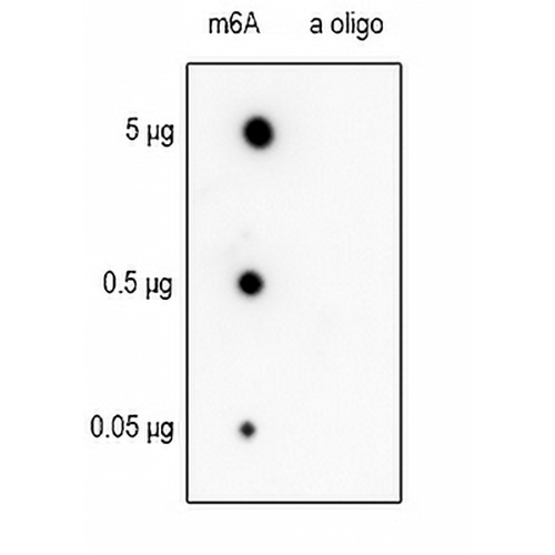
Key features and details
- Target:
- Source/Host:
- Reactivity:
- Clonality:
- Applications:
- Conjugation:
- Storage:
-
Brand:

 Product Details
DB | 1:500 - 1:2000 |
Description | Recombinant rabbit monoclonal antibody to N6-methyladenosine |
Specificity | Recognizes N6-methyladenosine |
Antibody Type | Primary antibody, Recombinant |
Imnunogen | KLH-conjugated m6A |
Purification | The antibody was purified by immunogen affinity chromatography. |
Molecular Weight | N/A |
Form/Buffer | Liquid in PBS, pH 7.3, 50% glycerol, 0.05% BSA, and 0.05% Proclin300. |
Alternative Names | m6A |
Gene Symbol | |
Entrez Gene | |
SwissProt |
*Clone Number, Reactivity, Source/Host and Clonality can be found in the product name and Key Features section above.
The membrane was blotted with anti-N6-methyladenosine antibody. The HRP-conjugated Goat anti-Rabbit IgG (H+L) antibody was used to detect the antibody.

Immunohistochemical analysis of Phospho-Serine/Threonine staining in human brain formalin fixed paraffin embedded tissue section. The section was pre-treated using heat mediated antigen retrieval with sodium citrate buffer (pH 6.0). The section was then incubated with the antibody at room temperature and detected using an HRP conjugated compact polymer system. DAB was used as the chromogen. The section was then counterstained with haematoxylin and mounted with DPX.

Immunofluorescent analysis of Phospho-Serine/Threonine staining in Hela cells. Formalin-fixed cells were permeabilized with 0.1% Triton X-100 in TBS for 5-10 minutes and blocked with 3% BSA-PBS for 30 minutes at room temperature. Cells were probed with the primary antibody in 3% BSA-PBS and incubated overnight at 4 °C in a humidified chamber. Cells were washed with PBST and incubated with an AREX® Fluor 488 -conjugated secondary antibody (green) in PBS at room temperature in the dark. Phalloidin - AREX® Fluor 594 was used to stain Actin filaments (red). DAPI was used to stain the cell nuclei (blue).
FAQs
 New Products