Myo D1 Rabbit Monoclonal Antibody(ARB860)
 Datasheet
Key features and details
- Target:
- Clone ID:
- Source/Host:
- Reactivity:
- Applications:
- Dilution:
- Clonality:
- Storage:
-
Brand:

CAT.NO. : ARB6652
US$ Please choose
US$ Please choose
Size:
Trail, Bulk size or Custom requests Please contact us
 Product Details
Background
MyoD1, one of the MyoD family of myogenic helix - loop - helix transcription factors, combined with myogenin, plays a role in coordinating the myogenic differentiation pathway from the determination of mesodermal precursors into myoblasts, the differentiation of myoblasts into myotubes, and finally the maturation of myotubes into skeletal myofibers. Normal mature skeletal muscle does not express MyoD1 protein. MyoD1 is expressed in myoblasts before differentiation while myogenin has post - differentiation functions. Anti - MyoD1 immunostaining identifies cells committed to myogenesis in their earliest phase, thus, it is a better biomarker for less differentiated Rhabdomyosarcoma cells (RMS). RMS are the most frequent malignant soft tissue neoplasms of childhood. While better differentiated RMS have cross - striations or rhabdomyoblasts that allow for a confident morphologic diagnosis, less differentiated RMS resemble other small blue round - cell tumors. Studies suggest, anti - MyoD1 may be used together with anti - myogenin and anti - desmin as a panel of markers since any RMS is virtually never negative for all three markers simultaneously.
Application
To ensure optimal assay performance, AREX recommends conducting reagent titration tailored to each testing system for optimal detection results.
*Results are sample-specific. Please refer to your local assay conditions and test parameters for reference.
Application | Dilution Ratio |
IHC | 1:100 - 1:200 |
Overview
Predicted Molecular Wt | 35 kDa |
Purity | ProA affinity purified IgG |
Subcellular location | Nucleus |
Swissprot ID | P15172 |
Immunogen | Synthetic peptide corresponding to residues within aa1-100 of Human MyoD1 |
Storage Buffer | PBS 59%, Sodium azide 0.01%, Glycerol 40%, BSA 0.05% |
Recommended method | Heat induced epitope retrieval with Tris-EDTA buffer (pH 9.0), primary antibody incubate at RT (18° C-25° C) for 30 minutes. |
*Clone Number, Reactivity, Source/Host and Clonality can be found in the product name and Key Features section above.
Data
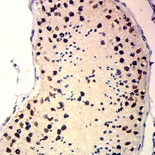
Immunohistochemistry analysis of Rhabdomyosarcoma tissue labeling Myo D1 with ARB860.
Storage
Store at 4°C short term. For long term storage, store at -20°C, avoiding freeze/thaw cycles.
Note
For Research Use Only. Not for diagnostic, therapeutics, prophylactic or in vivo use.
FAQs
Are the pathology antibodies provided by AREX raw antibodies or ready-to-use solutions?

AREX Biosciences specializes in supplying high-quality IHC pathology raw antibodies (Raw Antibodies). We do not manufacture ready-to-use working solutions or IVD diagnostic reagents. We mainly provide concentrated raw materials to pathology reagent manufacturers and diagnostic platform companies to support product development and OEM production.
What development scenarios are pathology raw antibodies mainly used for?
Our raw antibodies are primarily used for the research, performance optimization, validation, and commercial-scale production of pathology IHC detection reagents. They are also suitable for companion diagnostics (CDx) projects and antibody screening.
How can I evaluate whether a raw antibody is suitable for our staining platform?
It is recommended to focus on specificity, sensitivity, background control, and performance on your target automated platforms (such as Roche Ventana, Leica Bond, Agilent Dako, etc.). We can provide internal test data for reference, but we recommend partners perform actual validation on their own platforms.
Can you provide samples for platform validation?
Yes. We can provide validation samples depending on the specific product. Some products may be provided free of charge, while others may involve a small sample fee. Handling fee and shipping fee will be charged separately. Please contact us for detailed confirmation.
How should raw antibodies be paired with detection systems?
AREX raw antibodies can be used with various polymer detection systems and ancillary reagents. We recommend optimization in combination with the ARExVisual® system or your existing detection platform to achieve better signal intensity and background control.
New Products