L-Lactyl-Histone H3 (Lys18) Rabbit Monoclonal Antibody(ARA873)-ChIP Grade
 Datasheet
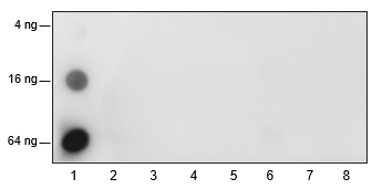
Key features and details
- Target:
- Host:
- Reactivity:
- Clonality:
- Application:
- Storage:
-
Brand:

 Product Details
Application | Dilution Ratio |
WB | 1:500 - 1:1000 |
ChIP | 6 μg/5×10⁶ cells |
Cut&tag | 1:100 |
Isotype | IgG |
Purification | Protein A |
Cellular Localization | Nucleus |
Form/Buffer | PBS, Glycerol, BSA |
UniProt ID | P68431 |
Synonyms | H3K18la |
Conjugate | Unconjugated |
Specificity | Anti-L-Lactyl-Histone H3 (Lys18) Rabbit mAb (ChIP Grade) detects histone H3 only when it is L-lactylated at Lys18. |
UniProt ID | P68431 |
Immunogen | L-lactylated human histone H3 (Lys18) peptide |
MW (kDa) | 15 |
*Clone Number, Reactivity, Source/Host and Clonality can be found in the product name and Key Features section above.
<p>Peptide amount: 4 ng, 16 ng, 64 ng <br>Blocking buffer: 5% NFDM/TBST<br>Primary Ab dilution: 1:2000 <br>Primary Ab incubation: 2 hours at room temperature<br>Secondary Ab: Goat Anti-Rabbit IgG H&L pAb (HRP Conjugate) <br>Exposure time: 10 seconds <br>The list of peptides used in the experiment is provided below.<br>Lane 1: H3K18 L-lactyl. Lane 2: H3K18 acetyl. <br>Lane 3: H3K18 crotonyl. Lane 4: H3K18 butyryl. <br>Lane 5: H3K18 propionyl. Lane 6: H3K18 2-hydroxyisobutyryl.<br>Lane 7: H3K18 β-hydroxybutyryl. Lane 8: H3K18 unmodified</p>
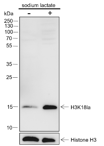
<p>Lysates: (-) HeLa cells; (+) HeLa cells treated with 25 mM sodium lactate for 24 hours<br>Protein loading amount: 20 μg <br>Blocking buffer: 5% NFDM/TBST <br>Primary Ab dilution: 1:1000 <br>Primary Ab incubation: 2 hours at room temperature <br>Secondary Ab: Goat Anti-Rabbit IgG H&L pAb (HRP Conjugate) <br>Exposure time: 60 seconds <br>Predicted band size: 15 kDa<br>Observed band size: 15 kDa</p>
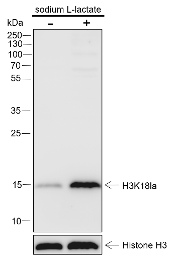
<p>Lysates: (-) K562 cells; (+) K562 cells treated with 25 mM sodium lactate for 24 hours <br>Protein loading amount: 20 μg <br>Blocking buffer: 5% NFDM/TBST <br>Primary Ab dilution: 1:1000 <br>Primary Ab incubation: 2 hours at room temperature <br>Secondary Ab: Goat Anti-Rabbit IgG H&L pAb (HRP Conjugate) <br>Exposure time: 60 seconds <br>Predicted band size: 15 kDa<br>Observed band size: 15 kDa</p>
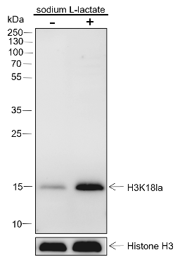
<p>Lysates: (-) NIH/3T3 cells; (+) NIH/3T3 cells treated with 25 mM sodium lactate for 24 hours<br>Protein loading amount: 20 μg <br>Blocking buffer: 5% NFDM/TBST <br>Primary Ab dilution: 1:1000 <br>Primary Ab incubation: 2 hours at room temperature <br>Secondary Ab: Goat Anti-Rabbit IgG H&L pAb (HRP Conjugate) <br>Exposure time: 60 seconds <br>Predicted band size: 15 kDa<br>Observed band size: 15 kDa</p>
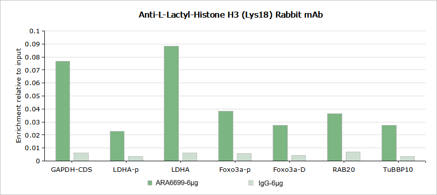
<p>Sample: HeLa cells treated with 100 mM sodium lactate for 24 hours<br>Cross-linking conditions: no cross-linking <br>Amount of chromatin per IP: 5×10<sup>6</sup> cells <br>Amount of Ab per IP: 6 μg <br>Beads type and amount per IP: 50 μL of Protein A MagBeads <br>Description: Chromatin immunoprecipitations were performed with 6 μg of normal rabbit IgG as a negative control. The immunoprecipitated DNA was quantified by real-time PCR using primers specific for the human GAPDH-CDS, LDHA promoter, LDHA, FOXO3a downstream, RAB20, and TUBBP10 regions. The data are presented as enrichment of each sample relative to the total amount of input at each amplicon.</p>
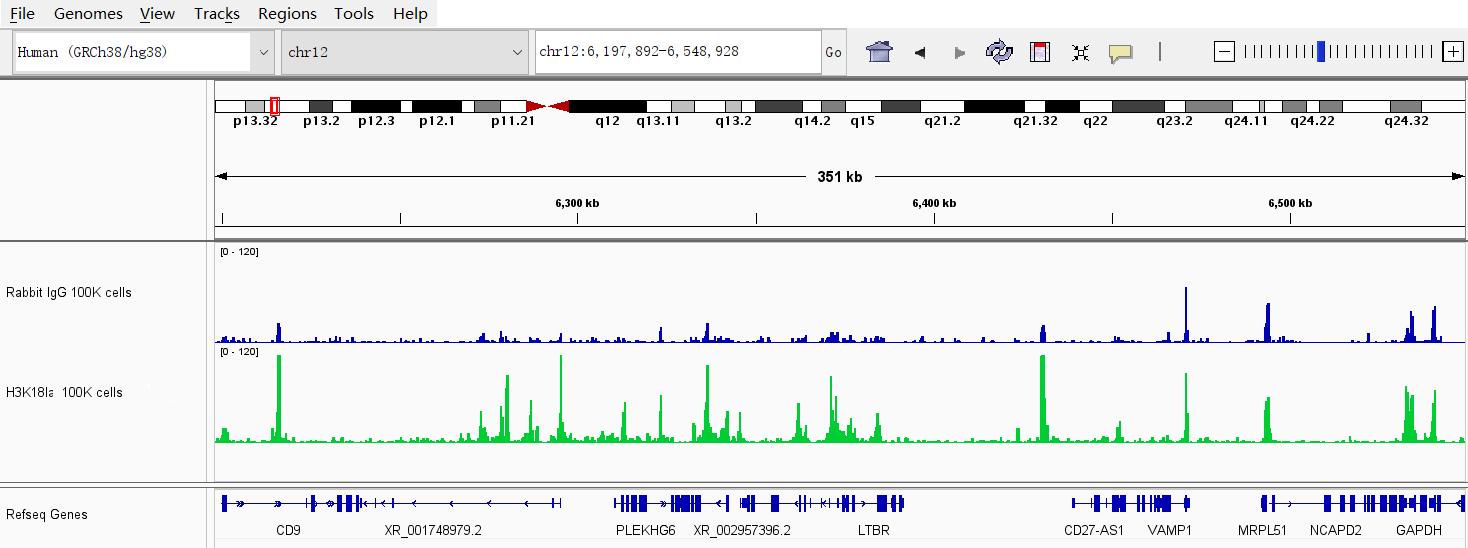
<p>Sample: HeLa cells treated with 100 mM sodium lactate for 24 hours<br>Cell quantity: 1×10<sup>5</sup> cells <br>Primary Ab dilution: 1:100 <br>Primary Ab incubation: 4⁰C overnight <br>Secondary Ab: Goat Anti-Rabbit IgG H&L pAb <br>Secondary Ab dilution: 1:1400 <br>Description: CUT&Tag was performed with Anti-L-Lactyl-Histone H3 (Lys18) Rabbit mAb alongside normal rabbit IgG as a negative control. The resulting DNA was sequenced on the Illumina NovaSeq platform with paired-end reads of 150 bp and a sequencing depth of 3 million reads. The figure shows significant H3K18la signal enrichments at the promoter regions of CD9, PLEKHG6, LTBR, and GAPDH.</p>
FAQs
 New Products