Histone H2B (Acetyl-K34) Rabbit Polyclonal Antibody
 MSDS
MSDS 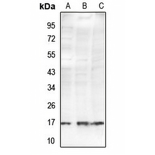
Key features and details
- Target:
- Source/Host:
- Reactivity:
- Clonality:
- Applications:
- Conjugation:
- Storage:
-
Brand:

 Product Details
WB | 1:500 - 1:1000 |
IF/ICC | 1:50 - 1:200 |
Description | Rabbit polyclonal antibody to Histone H2B (Acetyl-K34) |
Specificity | Recognizes endogenous levels of Histone H2B protein only when acetylated at K34. |
Antibody Type | Primary antibody |
Imnunogen | KLH-conjugated synthetic acetylated peptide corresponding to residues surrounding K34 of human Histone H2B protein. The exact sequence is proprietary. |
Purification | The antibody was purified by immunogen affinity chromatography. |
Molecular Weight | Predicted: 13 kD; Observed: 15 kD |
Form/Buffer | Liquid in 0.42% Potassium phosphate, 0.87% Sodium chloride, pH 7.3, 30% glycerol, and 0.01% sodium azide. |
Alternative Names | HIST1H2BC; H2BFL; HIST1H2BE; H2BFH; HIST1H2BF; H2BFG; HIST1H2BG; H2BFA; HIST1H2BI; H2BFK; Histone H2B type 1-C/E/F/G/I; Histone H2B.1 A; Histone H2B.a; H2B/a; Histone H2B.g; H2B/g; Histone H2B.h; H2B/h; Histone H2B.k; H2B/k; Histone H2B.l; H2B/l; HIST1H2BD; H2BFB; HIRIP2; Histone H2B type 1-D; HIRA-interacting protein 2; Histone H2B.1 B; Histone H2B.b; H2B/b; HIST1H2BH; H2BFJ; Histone H2B type 1-H; Histone H2B.j; H2B/j; HIST1H2BK; H2BFT; HIRIP1; Histone H2B type 1-K; H2B K; HIRA-interacting protein 1; HIST1H2BL; H2BFC; Histone H2B type 1-L; Histone H2B.c; H2B/c; HIST1H2BM; H2BFE; Histone H2B type 1-M; Histone H2B.e; H2B/e; HIST1H2BN; H2BFD; Histone H2B type 1-N; Histone H2B.d; H2B/d; HIST2H2BF; Histone H2B type 2-F; H2BFS; Histone H2B type F-S; Histone H2B.s; H2B/s |
Gene Symbol | HIST1H2BC; HIST1H2BE; HIST1H2BF; HIST1H2BG; HIST1H2BI; HIST1H2BD; HIST1H2BH; HIST1H2BK; HIST1H2BL; HIST1H2BM; HIST1H2BN; HIST2H2BF; H2BFS |
Entrez Gene | 3017; 8339; 8343; 8344; 8346; 8347; 3017; 8345; 85236; 8340; 8342; 8341; 440689(Human); 319179; 319182; 319184(Mouse) |
SwissProt | P62807; P58876; Q93079; O60814; Q99880; Q99879; Q99877; Q5QNW6; P57053(Human); Q6ZWY9; Q64478; Q8CGP1(Mouse) |
*Clone Number, Reactivity, Source/Host and Clonality can be found in the product name and Key Features section above.
Western blot analysis of Histone H2B (Acetyl-K34) expression in A549 (A), BV2 (B), PC12 (C) whole cell lysates. (Predicted band size: 13 kD; Observed band size: 15 kD)
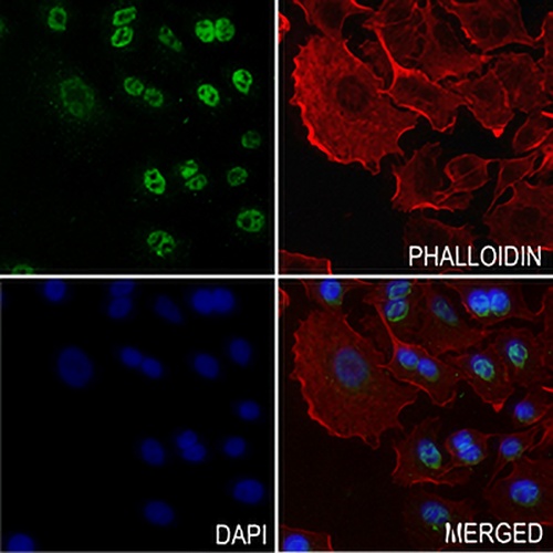
Immunofluorescent analysis of Histone H2B (Acetyl-K34) staining in A549 cells. Formalin-fixed cells were permeabilized with 0.1% Triton X-100 in TBS for 5-10 minutes and blocked with 3% BSA-PBS for 30 minutes at room temperature. Cells were probed with the primary antibody in 3% BSA-PBS and incubated overnight at 4 °C in a hidified chamber. Cells were washed with PBST and incubated with a AREX® Fluor 488 -conjugated secondary antibody (green) in PBS at room temperature in the dark. Phalloidin - AREX® Fluor 594 was used to stain Actin filaments (red). DAPI was used to stain the cell nuclei (blue).
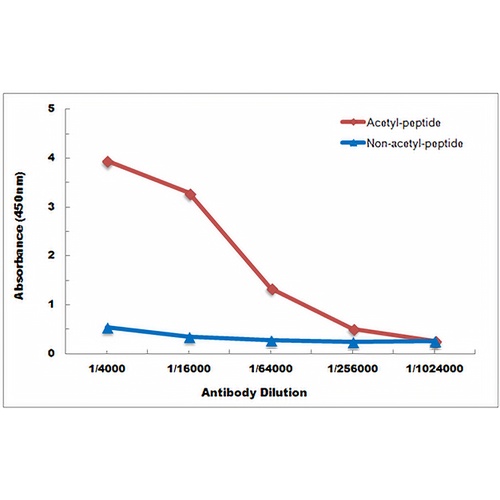
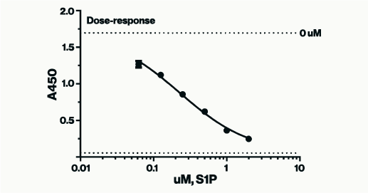
Direct ELISA antibody dose-response curve using Anti-Histone H2B (Acetyl-K34) Antibody. Antigen (Acetyl-peptide and non-acetyl-peptide) concentration is 5 ug/ml. Goat Anti-Rabbit IgG (H&L) - HRP was used as the secondary antibody, and signal was developed by TMB substrate.
FAQs
 New Products