HA tag Rabbit Monoclonal Antibody(ARA782)
 Datasheet
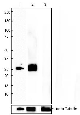
Key features and details
- Target:
- Source/Host:
- Reactivity:
- Clonality:
- Applications:
- Conjugation:
- Storage:
-
Brand:

 Product Details
WB | 1:1000-1:2000 |
IF/ICC | 1:400-1:2000 |
FC | 1:400-1:2000 |
IP | 1:10 |
Description | Rabbit Monoclonal Antibody to HA tag |
Antibody Type | Primary antibody |
Predicted MW | |
Immunogen | YPYDVPDYA (influenza hemagglutinin-HA-epitope) conjugated to KLH. |
Purification | ProA affinity purified IgG |
Form/Buffer | PBS 59%, Sodium azide 0.01%, Glycerol 40%, BSA 0.57%. |
Alternative Names | HA epitope tag; hemagglutinin |
*Clone Number, Reactivity, Source/Host and Clonality can be found in the product name and Key Features section above.
Predicted MW: Depend on fusion protein with HA tag
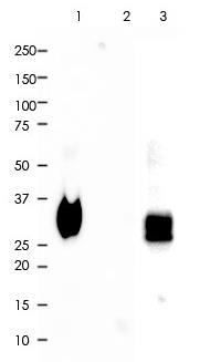
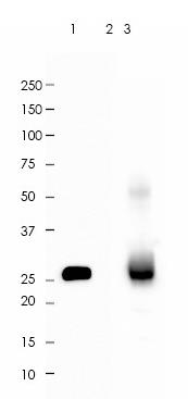
Lane 1: 293 cells lysate transfected with C-terminal HA tagged gene (ARA782 at 1:2,000 dilution).
Lane 2: 293 cells lysate transfected with N-terminal HA tagged gene (ARA782 at 1:1,000 dilution).
Lane 3: 293 cells lysate without any transfection (ARA782 at 1:400 dilution).
Lane 1: 1 µg per lane
Lane 2/3: 10 µg per lane
2nd Ab:
GAR HRP(H+L) 1:5,000
Exposure: 20s
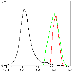
Overlay histogram showing 293 cells transfected with N-terminal (Green) and C-terminal (Red) HA tagged gene stained with ARA782. The cells were then incubated in the antibody (ARA782, 1:2,000 dilution) in 1x PBS/1% BSA for 30 min at room temperature. The secondary antibody used was a Goat Anti-Rabbit Alexa Fluor<sup>®</sup> 488 (IgG H+L) at 1:2,000 dilution for 20 min at room temperature. Unlabelled sample (Black) was used as a control.
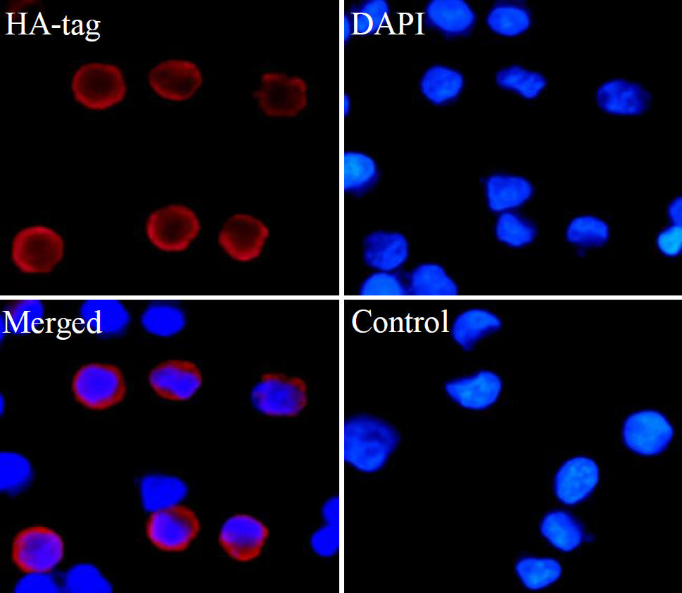
ARA782 staining HA tag in 293 cells transfected with N-terminal HA tagged gene by IF/ICC (immunofluorescence/immunocytochemistry). Cells were fixed with paraformaldehyde, permeabilized with 0.1% Triton X-100 and blocked with 10% goat serum for half an hour at room temperature. Samples were incubated with primary antibody (1:2,000) at 4°C. An Alexa Fluor<sup>®</sup> 594-conjugated Goat Anti-Rabbit IgG polyclonal was used as the secondary antibody (1:500). DAPI (blue) was used as the nuclear counter stain.
Control: PBS and secondary antibody, An Alexa Fluor<sup>®</sup> 594-conjugated Goat Anti-Rabbit IgG (1:500).
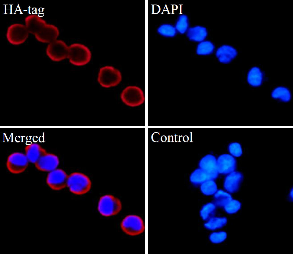
ARA782 staining HA tag in 293 cells transfected with C-terminal HA tagged gene by IF/ICC (immunofluorescence/immunocytochemistry). Cells were fixed with paraformaldehyde, permeabilized with 0.1% Triton X-100 and blocked with 10% goat serum for half an hour at room temperature. Samples were incubated with primary antibody (1:2,000) at 4°C. An Alexa Fluor<sup>®</sup> 594-conjugated Goat Anti-Rabbit IgG polyclonal was used as the secondary antibody (1:500). DAPI (blue) was used as the nuclear counter stain.
Control: PBS and secondary antibody, An Alexa Fluor<sup>®</sup> 594-conjugated Goat Anti-Rabbit IgG (1:500).
HA tag was immunoprecipitated from 0.2mg of 293 whole cells lysate transfected with N-terminal HA tagged gene with ARA782 at 1:10 dilution.
2nd Ab:
GAR HRP for IP 1:500
Lane 1: ARA782 IP in 293 whole cell lysate transfected with N-terminal HA tagged gene
Lane 2: PBS instead of ARA782 in 293 whole cell lysate transfected with N-terminal HA tagged gene
Lane 3: 293 whole cell lysate transfected with N-terminal HA tagged gene, 10 µg (input)
Exposure: 60s
HA tag was immunoprecipitated from 0.2mg of 293 whole cells lysate transfected with C-terminal HA tagged gene with ARA782 at 1:10 dilution.
2nd Ab:
GAR HRP for IP 1:500
Lane 1: ARA782 IP in 293 whole cell lysate transfected with C-terminal HA tagged gene
Lane 2: PBS instead of ARA782 in 293 whole cell lysate transfected with C-terminal HA tagged gene
Lane 3: 293 whole cell lysate transfected with C-terminal HA tagged gene, 10 µg (input)
Exposure: 60s
FAQs
 New Products