GPR56 Mouse Monoclonal Antibody(C4057)
 Datasheet
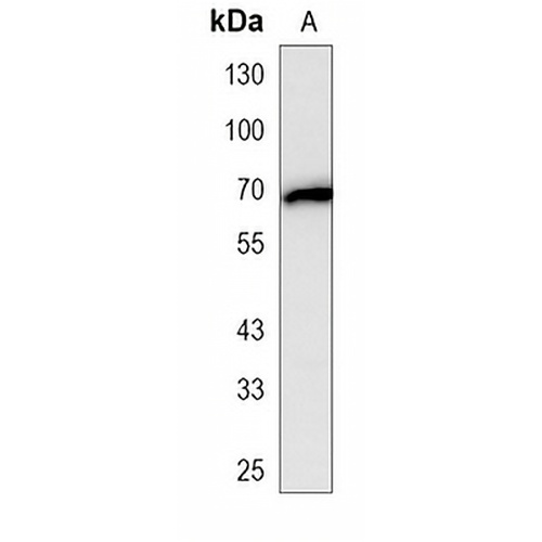
Key features and details
Mouse monoclonal antibody to GPR56
- Target:
- Source/Host:
- Reactivity:
- Clonality:
- Applications:
- Conjugation:
- Storage:
-
Brand:

CAT.NO. : AMA03669
US$ Please choose
US$ Please choose
 Product Details
Background
Adhesion G-protein coupled receptor (aGPCR) for steroid hormone 17alpha-hydroxypregnenolone (17-OH), which is involved in cell adhesion and cell-cell interactions for steroid hormone 17alpha-hydroxypregnenolone (17-OH), which is involved in cell adhesion and cell-cell interactions . Ligand binding causes a conformation change that triggers signaling via guanine nucleotide-binding proteins (G proteins) and modulates the activity of downstream effectors, such as RhoA pathway . ADGRG1 is coupled to G(12) and/or G(13) G proteins (GNA12 and GNA13, respectively) and mediates the activation Rho small GTPases . Acts as a potent suppressor of ferroptosis: binding to 17-OH-binding initiates signaling that down-regulates CD36 and alleviates ferroptosis-induced liver injury .
Application
To ensure optimal assay performance, AREX recommends conducting reagent titration tailored to each testing system for optimal detection results.
*Results are sample-specific. Please refer to your local assay conditions and test parameters for reference.
WB | 1:500 - 1:1000 |
Overview
Description | Mouse monoclonal antibody to GPR56 |
Specificity | Recognizes endogenous levels of GPR56 protein. |
Antibody Type | Primary antibody |
Imnunogen | Recombinant fusion protein of human GPR56. The exact sequence is proprietary. |
Purification | This antibody is purified through a protein G column. |
Molecular Weight | Predicted: 77 kD; Observed: 68 kD |
Form/Buffer | Mouse IgG1 kappa. Liquid in PBS, pH 7.3, 30% glycerol, and 0.01% sodium azide. |
Alternative Names | TM7LN4; TM7XN1; G-protein coupled receptor 56; Protein TM7XN1 |
Gene Symbol | GPR56 |
Entrez Gene | 9289(Human) |
SwissProt | Q9Y653(Human) |
*Clone Number, Reactivity, Source/Host and Clonality can be found in the product name and Key Features section above.
Data
Western blot analysis of GPR56 expression in Hela (A) whole cell lysates. (Predicted band size: 77 kD; Observed band size: 68 kD)
Storage
Store at 4°C short term. For long term storage, store at -20°C, avoiding freeze/thaw cycles.
Note
For Research Use Only. Not for diagnostic, therapeutics, prophylactic or in vivo use.
FAQs
What are the main types of research antibodies and how do they differ?

Research antibodies are mainly divided into monoclonal antibodies and polyclonal antibodies. Monoclonal antibodies typically offer higher specificity and better batch-to-batch consistency, while polyclonal antibodies often provide stronger affinity but may show more variation between batches. The choice depends on your specific experimental needs.
How can I tell if a research antibody is suitable for my experiment?
It is recommended to carefully review the product datasheet for validated applications, species reactivity, recommended dilutions, and published references. For new antibodies, performing a small-scale validation with positive control samples is usually helpful.
Can improper storage of research antibodies affect experimental results?
Yes. Antibodies are sensitive to temperature, repeated freeze-thaw cycles, and contamination. Improper storage may lead to reduced activity, increased background, or weaker signals. It is best to follow the storage instructions provided in the product datasheet.
Why doesn’t the recommended dilution in the datasheet work well in my experiment?
The recommended dilution is based on the supplier’s test conditions. Factors such as sample type, fixation method, and detection system in your lab can influence the optimal working concentration. Performing a dilution series optimization in your own system is often necessary.
What precautions should I take when using a newly purchased research antibody for the first time?
It is advisable to briefly centrifuge the antibody (especially concentrated or lyophilized ones), then perform a small-scale pilot experiment using the recommended conditions. Recording the batch number and usage date is also helpful for future tracking.
New Products