Cytokeratin 13 Mouse Monoclonal Antibody(C2260)

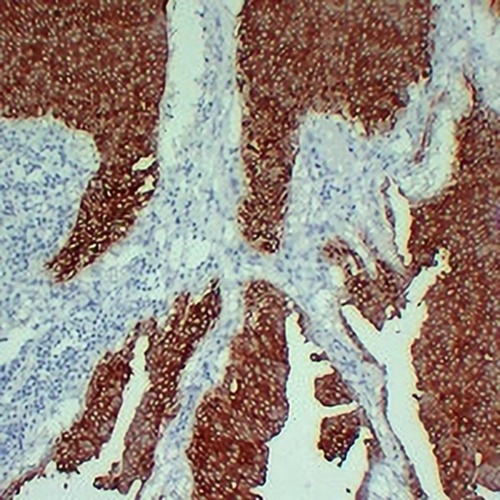
Key features and details

Mouse monoclonal antibody to Cytokeratin 13
  • Target: Cytokeratin 13
  • Source/Host: Mouse
  • Reactivity: Human
  • Clonality: Monoclonal
  • Applications: WB, IHC
  • Conjugation: Unconjugated
  • Storage: at-20°C
  • Brand:
CAT.NO. : AMA01872
US$ Please choose
US$ Please choose
Size:
Trail, Bulk size or Custom requests Please contact us
Product Details
Background
Type 1 keratin (Probable). Maintains postnatal tongue mucosal cell homeostasis and tissue organization in response to mechanical stress, potentially via regulation of the G1/S phase cyclins CCNE1 and CCNE2 .
Application
To ensure optimal assay performance, AREX recommends conducting reagent titration tailored to each testing system for optimal detection results.

WB

1:500 - 1:1000

IHC

1:100 - 1:300

*Results are sample-specific. Please refer to your local assay conditions and test parameters for reference.
Overview

Description

Mouse monoclonal antibody to Cytokeratin 13

Specificity

Recognizes endogenous levels of Cytokeratin 13 protein.

Antibody Type

Primary antibody

Imnunogen

KLH-conjugated synthetic peptide encompassing a sequence within human Cytokeratin 13. The exact sequence is proprietary.

Purification

The antibody was purified by immunogen affinity chromatography.

Molecular Weight

N/A

Form/Buffer

Mouse IgG1. Liquid in PBS containing 50% glycerol, 0.2% BSA and 0.01% sodium azide.

Alternative Names

Keratin type I cytoskeletal 13; Cytokeratin-13; CK-13; Keratin-13; K13

Gene Symbol

KRT13

Entrez Gene

3860(Human)

SwissProt

P13646(Human)

*AREX continuously optimizes our products. Webpage content may not reflect the latest updates. For inquiries, please contact info@arexbio.com or your local distributor.
*Clone Number, Reactivity, Source/Host and Clonality can be found in the product name and Key Features section above.
Data

Immunohistochemical analysis of Cytokeratin 13 staining in human transitional cell carcinoma formalin fixed paraffin embedded tissue section. The section was pre-treated using heat mediated antigen retrieval with sodium citrate buffer (pH 6.0). The section was then incubated with the antibody at room temperature and detected using an HRP conjugated compact polymer system. DAB was used as the chromogen. The section was then counterstained with haematoxylin and mounted with DPX.

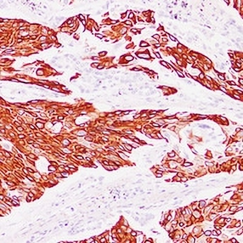
Immunohistochemical analysis of Cytokeratin 13 staining in human squamous cell lung carcinoma formalin fixed paraffin embedded tissue section. The section was pre-treated using heat mediated antigen retrieval with sodium citrate buffer (pH 6.0). The section was then incubated with the antibody at room temperature and detected using an HRP conjugated compact polymer system. DAB was used as the chromogen. The section was then counterstained with haematoxylin and mounted with DPX.

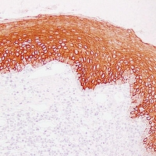
Immunohistochemical analysis of Cytokeratin 13 staining in human tonsil formalin fixed paraffin embedded tissue section. The section was pre-treated using heat mediated antigen retrieval with sodium citrate buffer (pH 6.0). The section was then incubated with the antibody at room temperature and detected using an HRP conjugated compact polymer system. DAB was used as the chromogen. The section was then counterstained with haematoxylin and mounted with DPX.

Storage
Store at 4°C short term. For long term storage, store at -20°C, avoiding freeze/thaw cycles.
Note
For Research Use Only. Not for use in diagnostic procedures.
FAQs
What are the main types of research antibodies and how do they differ?
Research antibodies are mainly divided into monoclonal antibodies and polyclonal antibodies. Monoclonal antibodies typically offer higher specificity and better batch-to-batch consistency, while polyclonal antibodies often provide stronger affinity but may show more variation between batches. The choice depends on your specific experimental needs.
How can I tell if a research antibody is suitable for my experiment?
It is recommended to carefully review the product datasheet for validated applications, species reactivity, recommended dilutions, and published references. For new antibodies, performing a small-scale validation with positive control samples is usually helpful.
Can improper storage of research antibodies affect experimental results?
Yes. Antibodies are sensitive to temperature, repeated freeze-thaw cycles, and contamination. Improper storage may lead to reduced activity, increased background, or weaker signals. It is best to follow the storage instructions provided in the product datasheet.
Why doesn’t the recommended dilution in the datasheet work well in my experiment?
The recommended dilution is based on the supplier’s test conditions. Factors such as sample type, fixation method, and detection system in your lab can influence the optimal working concentration. Performing a dilution series optimization in your own system is often necessary.
What precautions should I take when using a newly purchased research antibody for the first time?
It is advisable to briefly centrifuge the antibody (especially concentrated or lyophilized ones), then perform a small-scale pilot experiment using the recommended conditions. Recording the batch number and usage date is also helpful for future tracking.
New Products
Get in touch with AREX
Name:*
Tel/Phone:*
Company:*
Email:*
Inquiry:
Captcha*
Submitting your email information means that you are willing to receive email information from AREX regarding technology, applications, products, and events. By clicking on the 'unsubscribe' button in the email or by contacting info@arexbio.com You can unsubscribe at any time by sending an email. Regarding your data usage information, please refer to our privacy policy.
© AREX 2024. All Rights Reserved. Sitemap
0.505036s