CD167a Mouse Monoclonal Antibody(C3881)
 Datasheet
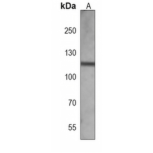
Key features and details
- Target:
- Source/Host:
- Reactivity:
- Clonality:
- Applications:
- Conjugation:
- Storage:
-
Brand:

 Product Details
WB | 1:500 - 1:1000 |
IHC | 1:50 - 1:200 |
Description | Mouse monoclonal antibody to CD167a |
Specificity | Recognizes endogenous levels of CD167a protein. |
Antibody Type | Primary antibody |
Imnunogen | Recombinant fusion protein of human CD167a. The exact sequence is proprietary. |
Purification | This antibody is purified through a protein G column. |
Molecular Weight | Predicted: 101 kD; Observed: 125 kD |
Form/Buffer | Mouse IgG2b kappa. Liquid in PBS, pH 7.3, 30% glycerol, and 0.01% sodium azide. |
Alternative Names | CAK; EDDR1; NEP; NTRK4; PTK3A; RTK6; TRKE; Epithelial discoidin domain-containing receptor 1; Epithelial discoidin domain receptor 1; CD167 antigen-like family member A; Cell adhesion kinase; Discoidin receptor tyrosine kinase; HGK2; Mammary carcinoma kinase 10; MCK-10; Protein-tyrosine kinase 3A; Protein-tyrosine kinase RTK-6; TRK E; Tyrosine kinase DDR; Tyrosine-protein kinase CAK; CD167a |
Gene Symbol | DDR1 |
Entrez Gene | 780(Human) |
SwissProt | Q08345(Human) |
*Clone Number, Reactivity, Source/Host and Clonality can be found in the product name and Key Features section above.
Western blot analysis of CD167a expression in MCF7 (A) whole cell lysates. (Predicted band size: 101 kD; Observed band size: 125 kD)
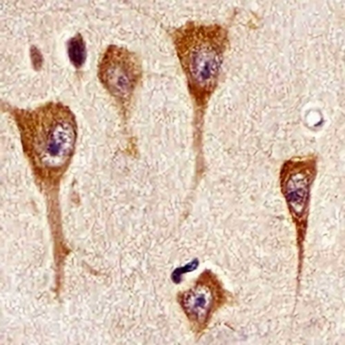
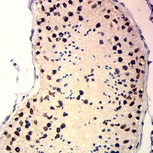
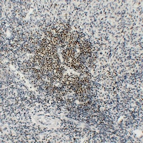
Immunohistochemical analysis of CD167a staining in human brain formalin fixed paraffin embedded tissue section. The section was pre-treated using heat mediated antigen retrieval with sodium citrate buffer (pH 6.0). The section was then incubated with the antibody at room temperature and detected using an HRP conjugated compact polymer system. DAB was used as the chromogen. The section was then counterstained with haematoxylin and mounted with DPX.
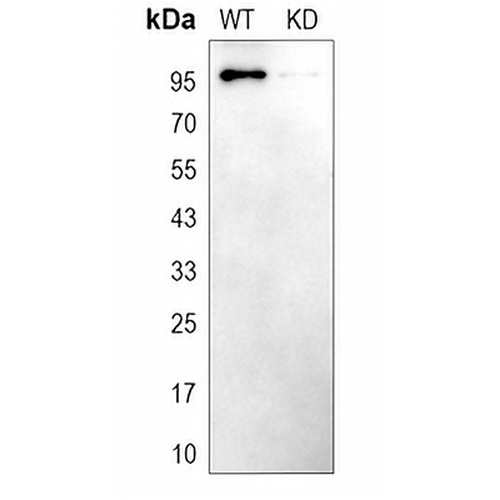
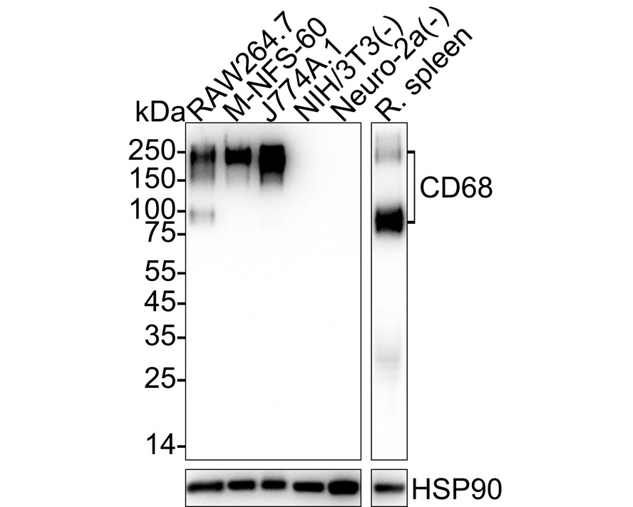
Western blot analysis of CD167a expression in wild type (WT) and knockdown (KD) HeLa cell lysates.
FAQs
 New Products