c-Jun Rabbit Polyclonal Antibody
 MSDS
MSDS 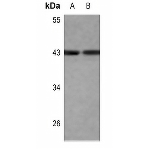
Key features and details
- Target:
- Source/Host:
- Reactivity:
- Clonality:
- Applications:
- Conjugation:
- Storage:
-
Brand:

 Product Details
WB | 1:500 - 1:1000 |
IHC | 1:100 - 1:200 |
IF/ICC | 1:50 - 1:200 |
IP | 1:10 - 1:100 |
FC | 1:100 - 1:300 |
Description | Rabbit polyclonal antibody to c-Jun |
Specificity | Recognizes endogenous levels of c-Jun protein. |
Antibody Type | Primary antibody |
Imnunogen | KLH-conjugated synthetic peptide encompassing a sequence within the center region of human c-Jun. The exact sequence is proprietary. |
Purification | The antibody was purified by immunogen affinity chromatography. |
Molecular Weight | Predicted: 35 kD; Observed: 43 kD |
Form/Buffer | Liquid in 0.42% Potassium phosphate, 0.87% Sodium chloride, pH 7.3, 30% glycerol, and 0.01% sodium azide. |
Alternative Names | Transcription factor AP-1; Activator protein 1; AP1; Proto-oncogene c-Jun; V-jun avian sarcoma virus 17 oncogene homolog; p39 |
Gene Symbol | JUN |
Entrez Gene | 3725(Human); 16476(Mouse); 24516(Rat) |
SwissProt | P05412(Human); P05627(Mouse); P17325(Rat) |
*Clone Number, Reactivity, Source/Host and Clonality can be found in the product name and Key Features section above.
Western blot analysis of c-Jun expression in PC3 (A), H1688 (B) whole cell lysates. (Predicted band size: 35 kD; Observed band size: 43 kD)
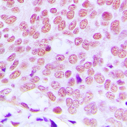
Immunohistochemical analysis of c-Jun staining in human breast cancer formalin fixed paraffin embedded tissue section. The section was pre-treated using heat mediated antigen retrieval with sodium citrate buffer (pH 6.0). The section was then incubated with the antibody at room temperature and detected using an HRP conjugated compact polymer system. DAB was used as the chromogen. The section was then counterstained with haematoxylin and mounted with DPX.
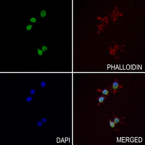
Immunofluorescent analysis of c-Jun staining in RAW264.7 cells. Formalin-fixed cells were permeabilized with 0.1% Triton X-100 in TBS for 5-10 minutes and blocked with 3% BSA-PBS for 30 minutes at room temperature. Cells were probed with the primary antibody in 3% BSA-PBS and incubated overnight at 4 °C in a hidified chamber. Cells were washed with PBST and incubated with a AREX® Fluor 488 -conjugated secondary antibody (green) in PBS at room temperature in the dark. Phalloidin - AREX® Fluor 594 was used to stain Actin filaments (red). DAPI was used to stain the cell nuclei (blue).
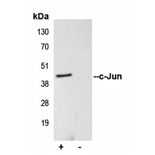
Immunoprecipitation of c-Jun from 0.5mg HEK293T whole cell extract lysate, using 5ug of Anti-c-Jun Antibody and 50ul of protein G magnetic beads (+). No antibody was added to the control (-). The antibody was incubated under agitation with Protein G beads for 10min, HEK293T whole cell extract lysate diluted in RIPA buffer was added to each sample and incubated for a further 10min under agitation. Proteins were eluted by addition of 40ul SDS loading buffer and incubated for 10min at 70°C; 10ul of each sample was separated on a SDS PAGE gel, transferred to a nitrocellulose membrane, blocked with 5% BSA and probed with Anti-c-Jun Antibody.
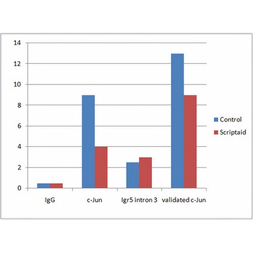
ChIP analysis of Human endothelial cells (EA.hy926), incubated for 10 hours at 4°C. Cross-linking (X-ChIP) using formaldehyde for 10 minutes. Positive control: Position 89340150-89340297 in chromosome 11 (has a validated c-Jun site). Negative Control: Igr5 intron 3 (contains no c-Jun binding site). Detection step: Real-time PCR.
FAQs
 New Products