Anti-Serotype 4 Fowl Adenovirus (FAdV-4) Fiber-2 Protein Mouse Monoclonal Antibody (1C9)
 Datasheet
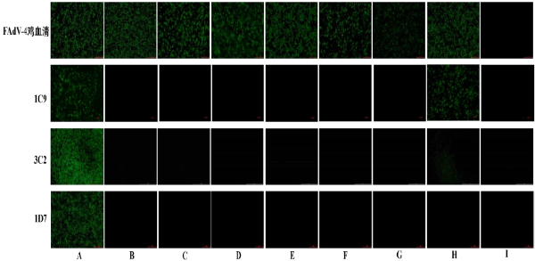
Key features and details
- Target:
- Host:
- Reactivity:
- Clonality:
- Application:
- Storage
-
Brand:

CAT.NO. : AXA0025
US$ Please choose
US$ Please choose
Size:
Trail, Bulk size or Custom requests Please contact us
 Product Details
Background
Serotype 4 Fowl Adenovirus (FAdV-4) Fiber-2 Protein is a structural glycoprotein located on the viral capsid surface alongside Fiber-1 it plays a complementary role in viral infection by mediating interactions with host cell receptors that may differ from those targeted by Fiber-1 it possesses unique immunogenic epitopes contributing to the overall immune response against FAdV-4 infection and exhibits distinct structural and functional characteristics compared to Fiber-1 of the same serotype as a specific viral component it serves as a valuable target for the development of serological diagnostic tools and subunit vaccines enhancing the specificity and efficacy of FAdV-4 detection and prevention it also provides insights into the viral tropism and pathogenic mechanisms of FAdV-4 through studies on its binding specificity and biological functions.
Application
To ensure optimal assay performance, AREX recommends conducting reagent titration tailored to each testing system for optimal detection results.
*Results are sample-specific. Please refer to your local assay conditions and test parameters for reference.
Application | Dilution Ratio |
IFA | 1:500-1:1000 |
WB | 1:2000 - 1:5000 |
IP | 1:1000 |
Overview
Description | Mouse Monoclonal antibody to Serotype 4 Fowl Adenovirus (FAdV-4) Fiber-2 Protein |
Antibody Type | Primary antibody |
Isotype | IgG |
Immunogen | Prokaryotic expression product GST-Fiber-2 of FAdV-4 Fiber-2 gene |
Form/Buffer | PBS, 20% Glycerol; preservative: 0.05% Sodium Azide |
Alternative Names | FAdV-4 Fiber-2, FAdV4 fiber 2, Fowl adenovirus 4 fiber-2 protein, FAdV-C fiber-2 |
*Clone Number, Reactivity, Source/Host and Clonality can be found in the product name and Key Features section above.
Data
The IF/ICC results showed that clone 1C9 can effectively recognize LMH cells infected with FAdV-4 and FAdV-10.
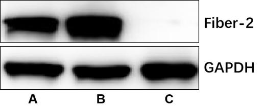
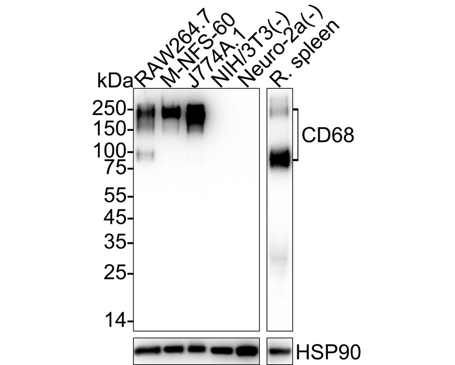
The WB results showed that clone 1C9 can effectively recognize the Fiber-2 protein of FAdV-4 and FAdV-10.
Reference
1. Wang P, Zhang J, Wang W, et al. A novel monoclonal antibody efficiently blocks the infection of serotype 4 fowl adenovirus by targeting fiber-2. Vet Res. 2018;49(1):29. doi:10.1186/s13567-018-0525-y
2. Xie S, Zhang J, Chen H, et al. Development of colloidal gold-based test strip for rapid detection of serotype 4 fowl adenovirus. J Virol Methods. 2021;296:114231. doi:10.1016/j.jviromet.2021.114231
3. Xie Q, Wang W, Kan Q, et al. FAdV-4 without Fiber-2 Is a Highly Attenuated and Protective Vaccine Candidate. Microbiol Spectr. 2022;10(1):e0143621. doi:10.1128/spectrum.01436-21
4. Xie Q, Cao S, Zhang W, et al. A novel fiber-2-edited live attenuated vaccine candidate against the highly pathogenic serotype 4 fowl adenovirus. Vet Res. 2021;52(1):35. Published 2021 Feb 27. doi:10.1186/s13567-021-00907-z
5. Xie Q, Wang W, Li L, et al. Domain in Fiber-2 interacted with KPNA3/4 significantly affects the replication and pathogenicity of the highly pathogenic FAdV-4. Virulence. 2021;12(1):754-765. doi:10.1080/21505594.2021.1888458
6. Mu Y, Xie Q, Wang W, et al. A Novel Fiber-1-Edited and Highly Attenuated Recombinant Serotype 4 Fowl Adenovirus Confers Efficient Protection Against Lethal Challenge. Front Vet Sci. 2021;8:759418. doi:10.3389/fvets.2021.759418
7. Wang W, Liu Q, Li T, et al. Fiber-1, Not Fiber-2, Directly Mediates the Infection of the Pathogenic Serotype 4 Fowl Adenovirus via Its Shaft and Knob Domains. J Virol. 2020;94(17):e00954-20. doi:10.1128/JVI.00954-20
Storage
Shipped at 4℃. Store at -20℃ for one year. Avoid repeated freeze/thaw cycles.
Note
For Research Use Only. Not for diagnostic, therapeutics, prophylactic or in vivo use.
FAQs
What are the main types of research antibodies and how do they differ?

Research antibodies are mainly divided into monoclonal antibodies and polyclonal antibodies. Monoclonal antibodies typically offer higher specificity and better batch-to-batch consistency, while polyclonal antibodies often provide stronger affinity but may show more variation between batches. The choice depends on your specific experimental needs.
How can I tell if a research antibody is suitable for my experiment?
It is recommended to carefully review the product datasheet for validated applications, species reactivity, recommended dilutions, and published references. For new antibodies, performing a small-scale validation with positive control samples is usually helpful.
Can improper storage of research antibodies affect experimental results?
Yes. Antibodies are sensitive to temperature, repeated freeze-thaw cycles, and contamination. Improper storage may lead to reduced activity, increased background, or weaker signals. It is best to follow the storage instructions provided in the product datasheet.
Why doesn’t the recommended dilution in the datasheet work well in my experiment?
The recommended dilution is based on the supplier’s test conditions. Factors such as sample type, fixation method, and detection system in your lab can influence the optimal working concentration. Performing a dilution series optimization in your own system is often necessary.
What precautions should I take when using a newly purchased research antibody for the first time?
It is advisable to briefly centrifuge the antibody (especially concentrated or lyophilized ones), then perform a small-scale pilot experiment using the recommended conditions. Recording the batch number and usage date is also helpful for future tracking.
New Products