Anticorpo policlonale di coniglio EMP
 Scheda tecnica
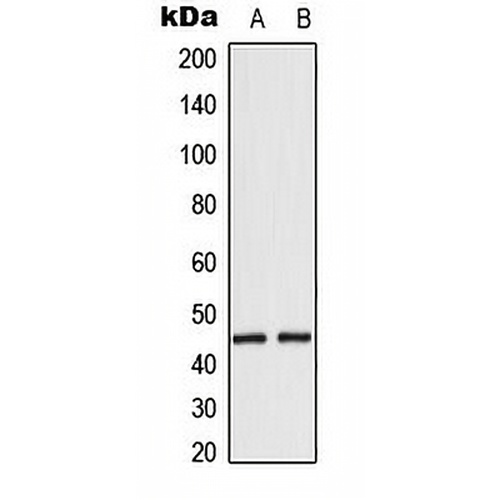
Caratteristiche principali e dettagli
- Obiettivo:
- Fonte/Ospite:
- Reattività:
- Clonalità:
- Applicazioni:
- Coniugazione:
- Stoccaggio:
-
Marca:

 Dettagli del prodotto
WB | 1:500 - 1:1000 |
IHC | 1:100 - 1:200 |
SE/ICC | 1:50 - 1:200 |
Descrizione | Anticorpo policlonale di coniglio anti-EMP |
Specificità | Riconosce i livelli endogeni della proteina EMP. |
Tipo di anticorpi | Anticorpo primario |
Immunogeno | KLH-conjugated synthetic peptide encompassing a sequence within the center region of human EMP. The exact sequence is proprietary. |
Purificazione | L'anticorpo è stato purificato mediante cromatografia di affinità immunogena. |
Peso Molecolare | Previsto: 45 kD; Osservato: 45 kD |
Modulo/Buffer | Liquid in 0.42% Potassium phosphate, 0.87% Sodium chloride, pH 7.3, 30% glycerol, and 0.01% sodium azide. |
Nomi alternativi | EMP; Macrophage erythroblast attacher; Cell proliferation-inducing gene 5 protein; Erythroblast macrophage protein; Human lung cancer oncogene 10 protein; HLC-10 |
Simbolo del gene | MAEA |
Entrez Gene | 10296(Umano); 59003(Topo); 298982(Ratto) |
SwissProt | Q7L5Y9(Umano); Q4VC33(mouse); Q5RKJ1(Ratto) |
*Clone Number, Reactivity, Source/Host and Clonality can be found in the product name and Key Features section above.
Western blot analysis of EMP expression in HepG2 (A), HeLa (B) whole cell lysates. (Predicted band size: 45 kD; Observed band size: 45 kD)
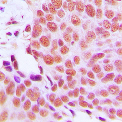
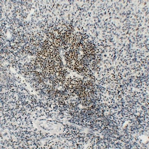
Immunohistochemical analysis of EMP staining in human breast cancer formalin fixed paraffin embedded tissue section. The section was pre-treated using heat mediated antigen retrieval with sodium citrate buffer (pH 6.0). The section was then incubated with the antibody at room temperature and detected using an HRP conjugated compact polymer system. DAB was used as the chromogen. The section was then counterstained with haematoxylin and mounted with DPX.
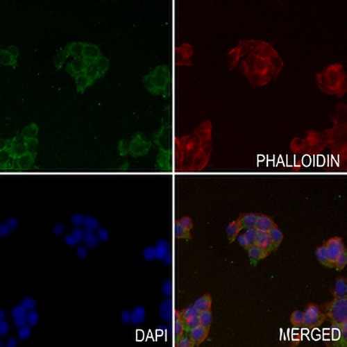
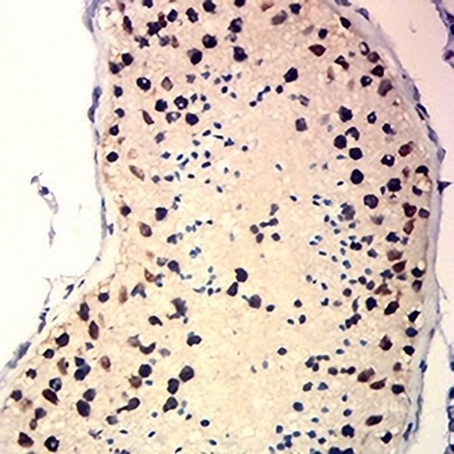
Immunofluorescent analysis of EMP staining in HEPG2 cells. Formalin-fixed cells were permeabilized with 0.1% Triton X-100 in TBS for 5-10 minutes and blocked with 3% BSA-PBS for 30 minutes at room temperature. Cells were probed with the primary antibody in 3% BSA-PBS and incubated overnight at 4 °C in a hidified chamber. Cells were washed with PBST and incubated with a AREX® Fluor 488 -conjugated secondary antibody (green) in PBS at room temperature in the dark. Phalloidin - AREX® Fluor 594 was used to stain Actin filaments (red). DAPI was used to stain the cell nuclei (blue).
Domande frequenti
 Nuovi prodotti