


Anticorpo policlonale di coniglio DNA-PKcs (Phospho-T2647).
 Scheda tecnica
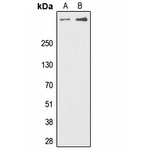
Caratteristiche principali e dettagli
- Obiettivo:
- Fonte/Ospite:
- Reattività:
- Clonalità:
- Applicazioni:
- Coniugazione:
- Stoccaggio:
-
Marca:

 Dettagli del prodotto
WB | 1:500 - 1:1000 |
IHC | 1:100 - 1:200 |
SE/ICC | 1:100 - 1:500 |
Descrizione | Anticorpo policlonale di coniglio contro DNA-PKcs (Phospho-T2647) |
Specificità | Riconosce i livelli endogeni della proteina DNA-PKcs solo quando fosforilata a T2647. |
Tipo di anticorpo | Anticorpo primario |
Immunogeno | KLH-conjugated synthetic phosphopeptide corresponding to residues surrounding T2647 of human DNA-PKcs protein. The exact sequence is proprietary. |
Purificazione | L'anticorpo è stato purificato mediante cromatografia di affinità immunogena. |
Peso Molecolare | Previsto: 469 kD; Osservato: 460 kD |
Modulo/Buffer | Liquid in 0.42% Potassium phosphate, 0.87% Sodium chloride, pH 7.3, 30% glycerol, and 0.01% sodium azide. |
Nomi alternativi | HYRC; HYRC1; DNA-dependent protein kinase catalytic subunit; DNA-PK catalytic subunit; DNA-PKcs; DNPK1; p460 |
Simbolo del gene | PRKDC |
Entrez Gene | 5591(Umano) |
SwissProt | P78527(Umano) |
*Clone Number, Reactivity, Source/Host and Clonality can be found in the product name and Key Features section above.
Western blot analysis of DNA-PKcs (Phospho-T2647) expression in Jurkat (A), HeLa (B) whole cell lysates. (Predicted band size: 469 kD; Observed band size: 460 kD)
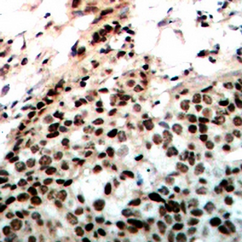
Immunohistochemical analysis of DNA-PKcs (Phospho-T2647) staining in human breast cancer formalin fixed paraffin embedded tissue section. The section was pre-treated using heat mediated antigen retrieval with sodium citrate buffer (pH 6.0). The section was then incubated with the antibody at room temperature and detected using an HRP conjugated compact polymer system. DAB was used as the chromogen. The section was then counterstained with haematoxylin and mounted with DPX.
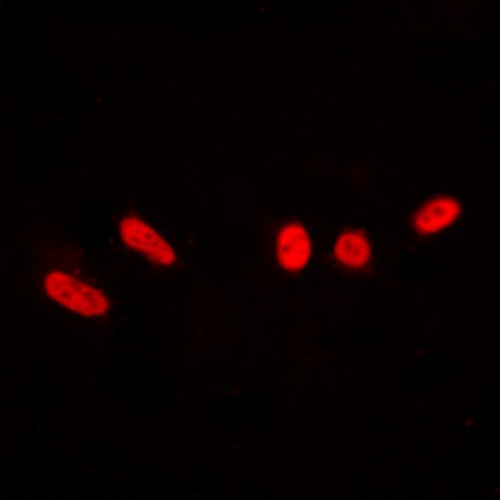
Immunofluorescent analysis of DNA-PKcs (Phospho-T2647) staining in Jurkat cells. Formalin-fixed cells were permeabilized with 0.1% Triton X-100 in TBS for 5-10 minutes and blocked with 3% BSA-PBS for 30 minutes at room temperature. Cells were probed with the primary antibody in 3% BSA-PBS and incubated overnight at 4 °C in a hidified chamber. Cells were washed with PBST and incubated with a DyLight 594-conjugated secondary antibody (red) in PBS at room temperature in the dark.
Domande frequenti
 Nuovi prodotti


