Anticuerpo policlonal de conejo PAK1 (fosfo-T212)
 Hoja de datos
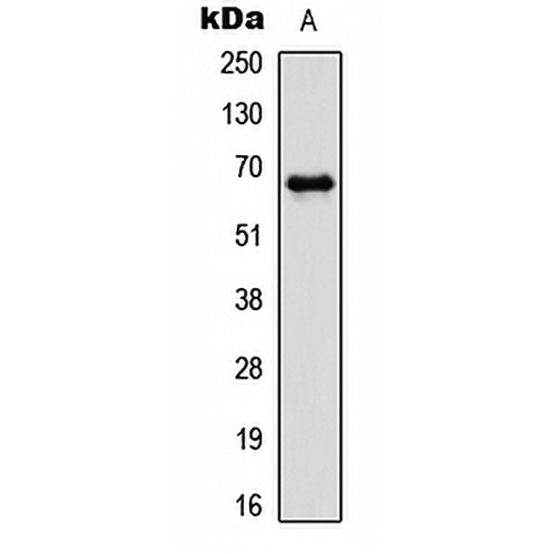
Características y detalles clave
- Objetivo:
- Fuente/Anfitrión:
- Reactividad:
- Clonalidad:
- Aplicaciones:
- Conjugación:
- Almacenamiento:
-
Marca:

 Detalles del producto
WB | 1:500 - 1:1000 |
IHC | 1:100 - 1:200 |
FI/CCI | 1:100 - 1:500 |
Descripción | Anticuerpo policlonal de conejo contra PAK1 (Phospho-T212) |
Especificidad | Reconoce niveles endógenos de proteína PAK1 solo cuando está fosforilada en T212. |
Tipo de anticuerpo | Anticuerpo primario |
Inmunógeno | KLH-conjugated synthetic phosphopeptide corresponding to residues surrounding T212 of human PAK1 protein. The exact sequence is proprietary. |
Purificación | El anticuerpo se purificó mediante cromatografía de afinidad de inmunógeno. |
Peso Molecular | Previsto: 60 kD; Observado: 68 kD |
Formulario/búfer | Liquid in 0.42% Potassium phosphate, 0.87% Sodium chloride, pH 7.3, 30% glycerol, and 0.01% sodium azide. |
Nombres alternativos | Serine/threonine-protein kinase PAK 1; Alpha-PAK; p21-activated kinase 1; PAK-1; p65-PAK |
Símbolo genético | PAK1 |
Entrez Gene | 5058 (humano); 29431 (Rata) |
SwissProt | Q13153 (Humano); O88643(Ratón); P35465 (Rata) |
*Clone Number, Reactivity, Source/Host and Clonality can be found in the product name and Key Features section above.
Western blot analysis of PAK1 (Phospho-T212) expression in HCT116 (A) whole cell lysates. (Predicted band size: 60 kD; Observed band size: 68 kD)
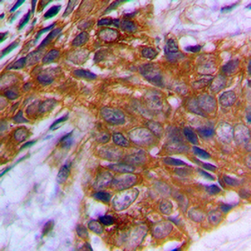
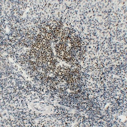
Immunohistochemical analysis of PAK1 (Phospho-T212) staining in human prostate cancer formalin fixed paraffin embedded tissue section. The section was pre-treated using heat mediated antigen retrieval with sodium citrate buffer (pH 6.0). The section was then incubated with the antibody at room temperature and detected using an HRP conjugated compact polymer system. DAB was used as the chromogen. The section was then counterstained with haematoxylin and mounted with DPX.
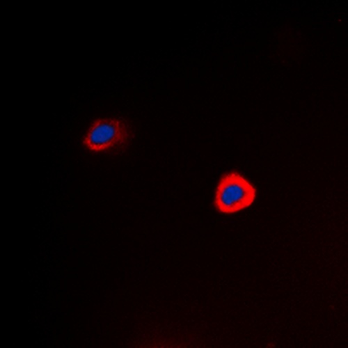
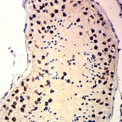
Immunofluorescent analysis of PAK1 (Phospho-T212) staining in Hela cells. Formalin-fixed cells were permeabilized with 0.1% Triton X-100 in TBS for 5-10 minutes and blocked with 3% BSA-PBS for 30 minutes at room temperature. Cells were probed with the primary antibody in 3% BSA-PBS and incubated overnight at 4 °C in a hidified chamber. Cells were washed with PBST and incubated with a DyLight 594-conjugated secondary antibody (red) in PBS at room temperature in the dark. DAPI was used to stain the cell nuclei (blue).
Preguntas frecuentes
 Nuevos productos