Anticuerpo policlonal de conejo NLRP3
 MSDS
MSDS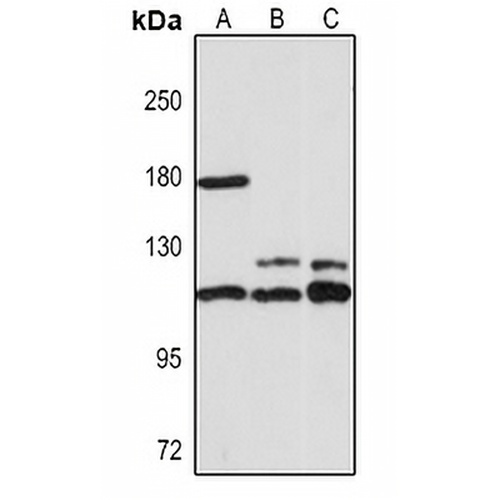
Características y detalles clave
- Objetivo:
- Fuente/Anfitrión:
- Reactividad:
- Clonalidad:
- Aplicaciones:
- Conjugación:
- Almacenamiento:
-
Marca:

 Detalles del producto
WB | 1:500 - 1:1000 |
IHC | 1:50 - 1:200 |
FI/CCI | 1:50 - 1:200 |
Descripción | Anticuerpo policlonal de conejo contra NLRP3 |
Especificidad | Reconoce niveles endógenos de la proteína NLRP3. |
Tipo de anticuerpo | Anticuerpo primario |
Inmunógeno | Proteína recombinante de NLRP3 humana. La secuencia exacta es propietaria. |
Purificación | El anticuerpo se purificó mediante cromatografía de afinidad de inmunógeno. |
Peso Molecular | Previsto: 83; Observado: 118 kD |
Formulario/búfer | Liquid in 0.42% Potassium phosphate, 0.87% Sodium chloride, pH 7.3, 30% glycerol, and 0.01% sodium azide. |
Nombres alternativos | C1orf7; CIAS1; NALP3; PYPAF1; NACHT, LRR and PYD domains-containing protein 3; Angiotensin/vasopressin receptor AII/AVP-like; Caterpiller protein 1.1; CLR1.1; Cold autoinflammatory syndrome 1 protein; Cryopyrin; PYRIN-containing APAF1-like protein 1 |
Símbolo genético | NLRP3 |
Entrez Gene | 114548 (humano); 216799 (ratón) |
SwissProt | Q96P20 (Humano); Q8R4B8 (ratón) |
*Clone Number, Reactivity, Source/Host and Clonality can be found in the product name and Key Features section above.
Western blot analysis of NLRP3 expression in HT29 (A), mouse brain (B), rat brain (C) whole cell lysates. (Predicted band size: 83; 105; 111; 112; 115; 118 kD; Observed band size: 118 kD)
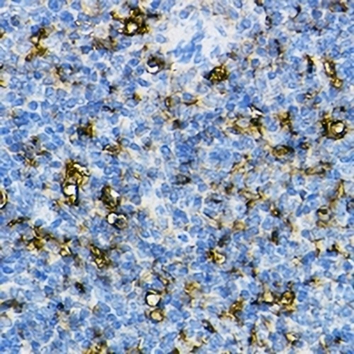
Immunohistochemical analysis of NLRP3 staining in mouse spleen formalin fixed paraffin embedded tissue section. The section was pre-treated using heat mediated antigen retrieval with sodium citrate buffer (pH 6.0). The section was then incubated with the antibody at room temperature and detected using an HRP conjugated compact polymer system. DAB was used as the chromogen. The section was then counterstained with haematoxylin and mounted with DPX.
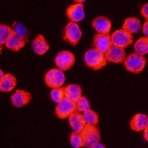
Immunofluorescent analysis of NLRP3 staining in Raw264.7 cells. Formalin-fixed cells were permeabilized with 0.1% Triton X-100 in TBS for 5-10 minutes and blocked with 3% BSA-PBS for 30 minutes at room temperature. Cells were probed with the primary antibody in 3% BSA-PBS and incubated overnight at 4 °C in a humidified chamber. Cells were washed with PBST and incubated with a DyLight 594-conjugated secondary antibody (red) in PBS at room temperature in the dark.
Preguntas frecuentes
 Nuevos productos