Anticuerpo monoclonal de ratón HSPA4 (C2752)
 MSDS
MSDS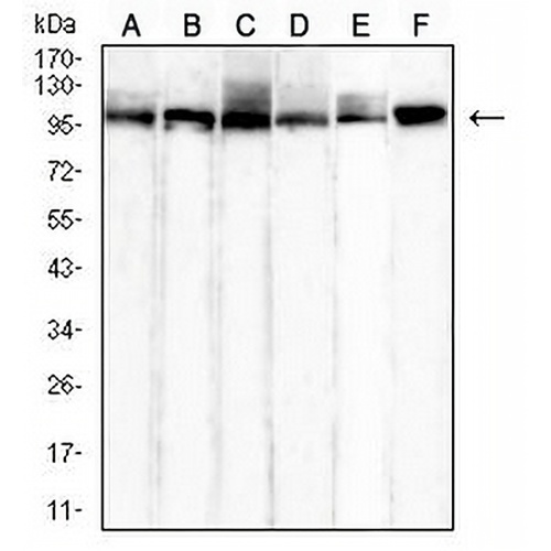
Características y detalles clave
- Objetivo:
- Fuente/Anfitrión:
- Reactividad:
- Clonalidad:
- Aplicaciones:
- Conjugación:
- Almacenamiento:
-
Marca:

 Detalles del producto
WB | 1:500 - 1:1000 |
IHC | 1:100 - 1:500 |
FI/CCI | 1:100 - 1:500 |
FC | 1:100 - 1:200 |
Descripción | Monoclonal de ratón para HSPA4 |
Especificidad | Reconoce niveles endógenos de proteína HSPA4. |
Tipo de anticuerpo | Anticuerpo primario |
Inmunógeno | Recombinant fusion protein of human HSPA4 expressed in HEK293 |
Purificación | Este anticuerpo se purifica a través de una columna de proteína G. |
Peso Molecular | Previsto: 94 kD; Observado: 100 kD kD |
Formulario/búfer | Mouse IgG1. Liquid in PBS, pH 7.3, 30% glycerol, and 0.01% sodium azide. |
Nombres alternativos | APG2; Heat shock 70 kDa protein 4; HSP70RY; Heat shock 70-related protein APG-2 |
Símbolo genético | HSPA4 |
Entrez Gene | 3308 (humano) |
SwissProt | P34932 (Humano) |
*Clone Number, Reactivity, Source/Host and Clonality can be found in the product name and Key Features section above.
Western blot analysis of HSPA4 expression in Hela (A), HepG2 (B), HEK293 (C), COS7 (D), A549 (E), Jurkat (F) whole cell lysates. (Predicted band size: 94 kD; Observed band size: 100 kD kD)
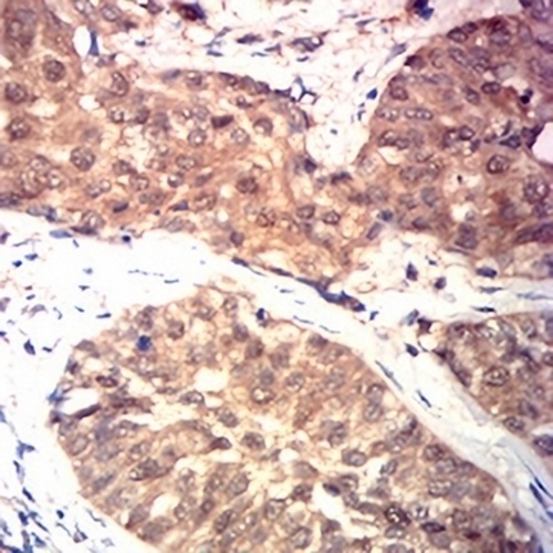
Immunohistochemical analysis of HSPA4 staining in human bladder cancer formalin fixed paraffin embedded tissue section. The section was pre-treated using heat mediated antigen retrieval with sodium citrate buffer (pH 6.0). The section was then incubated with the antibody at room temperature and detected using an HRP conjugated compact polymer system. DAB was used as the chromogen. The section was then counterstained with haematoxylin and mounted with DPX.
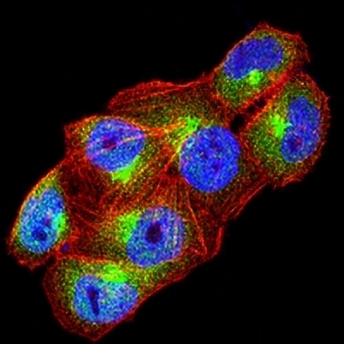
Immunofluorescent analysis of HSPA4 staining in Hela cells. Formalin-fixed cells were permeabilized with 0.1% Triton X-100 in TBS for 5-10 minutes and blocked with 3% BSA-PBS for 30 minutes at room temperature. Cells were probed with the primary antibody in 3% BSA-PBS and incubated overnight at 4 °C in a hidified chamber. Cells were washed with PBST and incubated with an AREX® Fluor 488 -conjugated secondary antibody (green) in PBS at room temperature in the dark. Phalloidin - AREX® Fluor 594 was used to stain Actin filaments (red). DAPI was used to stain the cell nuclei (blue).
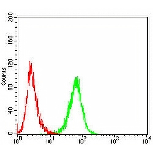
Flow cytometric analysis of Jurkat cells using Anti-HSPA4 Antibody (green) and negative control (red).
Preguntas frecuentes
 Nuevos productos