ERGIC-53 Rabbit Polyclonal Antibody
 MSDS
MSDS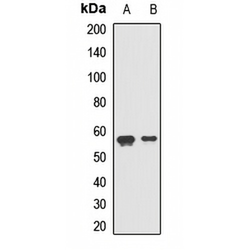
Características y detalles clave
- Objetivo:
- Fuente/Anfitrión:
- Reactividad:
- Clonalidad:
- Aplicaciones:
- Conjugación:
- Almacenamiento:
-
Marca:

 Detalles del producto
WB | 1:500 - 1:2000 |
FI/CCI | 1:50 - 1:200 |
Descripción | Rabbit polyclonal antibody to ERGIC-53 |
Especificidad | Recognizes endogenous levels of ERGIC-53 protein. |
Tipo de anticuerpo | Anticuerpo primario |
Inmunógeno | Recombinant fusion protein of human ERGIC-53 |
Purificación | The antibody was purified by immunogen affinity chromatography. |
Peso Molecular | Previsto: 57 kD; Observado: 57 kD |
Formulario/búfer | Liquid in 0.42% Potassium phosphate, 0.87% Sodium chloride, pH 7.3, 30% glycerol, and 0.01% sodium azide. |
Nombres alternativos | ERGIC53; F5F8D; Protein ERGIC-53; ER-Golgi intermediate compartment 53 kDa protein; Gp58; Intracellular mannose-specific lectin MR60; Lectin mannose-binding 1 |
Símbolo genético | LMAN1 |
Entrez Gene | 3998(Human); 70361(Mouse); 116666(Rat) |
SwissProt | P49257(Human); Q9D0F3(Mouse); Q62902(Rat) |
*Clone Number, Reactivity, Source/Host and Clonality can be found in the product name and Key Features section above.
Western blot analysis of ERGIC-53 expression in LO2 (A), U251 (B) whole cell lysates. (Predicted band size: 57 kD; Observed band size: 57 kD)
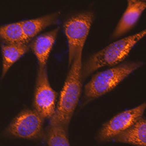
Immunofluorescent analysis of ERGIC-53 staining in NIH3T3 cells. Formalin-fixed cells were permeabilized with 0.1% Triton X-100 in TBS for 5-10 minutes and blocked with 3% BSA-PBS for 30 minutes at room temperature. Cells were probed with the primary antibody in 3% BSA-PBS and incubated overnight at 4 °C in a humidified chamber. Cells were washed with PBST and incubated with a AREX® Fluor 594-conjugated secondary antibody (red) in PBS at room temperature in the dark. DAPI was used to stain the cell nuclei (blue).
Preguntas frecuentes
 Nuevos productos