AKT (Phospho-Y315) Polyklonaler Kaninchen-Antikörper
 Datenblatt
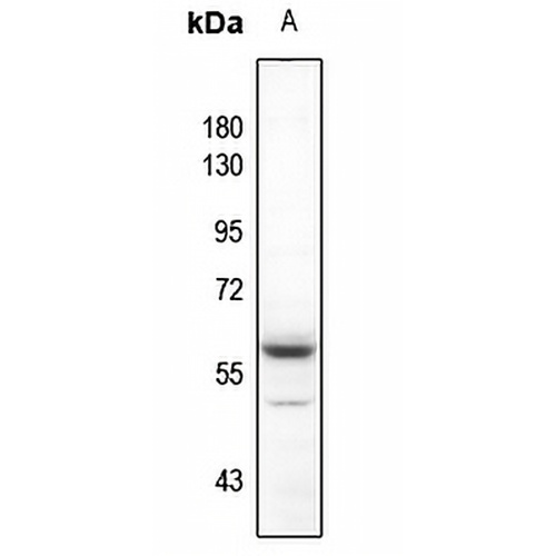
Hauptmerkmale und Details
- Ziel:
- Quelle/Host:
- Reaktivität:
- Klonalität:
- Anwendungen:
- Konjugation:
- Lagerung:
-
Marke:

 Produktdetails
WB | 1:500 - 1:1000 |
IHC | 1:100 - 1:200 |
IF/ICC | 1:100 - 1:500 |
Beschreibung | Polyklonaler Kaninchen-Antikörper gegen AKT (Phospho-Y315) |
Spezifität | Erkennt endogene Mengen an AKT-Protein nur, wenn es an Y315 phosphoryliert ist. |
Antikörpertyp | Primärer Antikörper |
Immunogen | KLH-conjugated synthetic phosphopeptide corresponding to residues surrounding Y315 of human AKT protein. The exact sequence is proprietary. |
Reinigung | Der Antikörper wurde durch Immunogenaffinitätschromatographie gereinigt. |
Molekulargewicht | Voraussichtlich: 55 kD; Beobachtet: 60 kD |
Form/Puffer | Flüssigkeit aus 0,42 % Kaliumphosphat, 0,87 % Natriumchlorid, pH 7,3, 30 % Glycerin und 0,01 % Natriumazid. |
Alternative Namen | AKT1; PKB; RAC; RAC-alpha serine/threonine-protein kinase; Protein kinase B; PKB; Protein kinase B alpha; PKB alpha; Proto-oncogene c-Akt; RAC-PK-alpha; AKT2; RAC-beta serine/threonine-protein kinase; Protein kinase Akt-2; Protein kinase B beta; PKB beta; RAC-PK-beta; AKT3; PKBG; RAC-gamma serine/threonine-protein kinase; Protein kinase Akt-3; Protein kinase B gamma; PKB gamma; RAC-PK-gamma; STK-2 |
Gensymbol | AKT1; AKT2; AKT3 |
Entrez Gene | 207; 208; 10000 (Mensch); 11651; 11652; 23797(Maus); 24185; 25233; 29414(Ratte) |
SwissProt | P31749; P31751; Q9Y243 (Mensch); P31750; Q60823; Q9WUA6(Maus); P47196; P47197; Q63484(Ratte) |
*Klonnummer, Reaktivität, Quelle/Host und Klonalität finden Sie oben im Abschnitt Produktname und Hauptfunktionen.
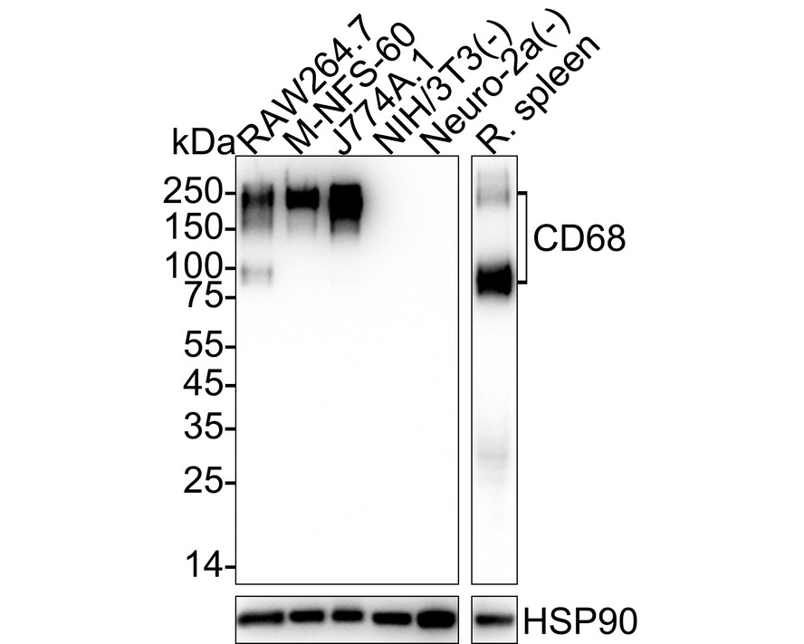
Western blot analysis of AKT (Phospho-Y315) expression in U87MG (A) whole cell lysates. (Predicted band size: 55 kD; Observed band size: 60 kD)
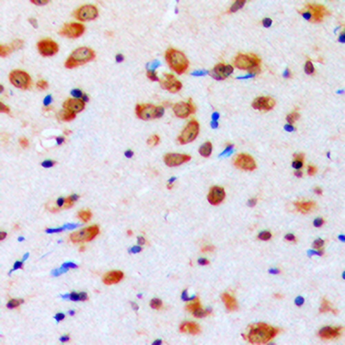
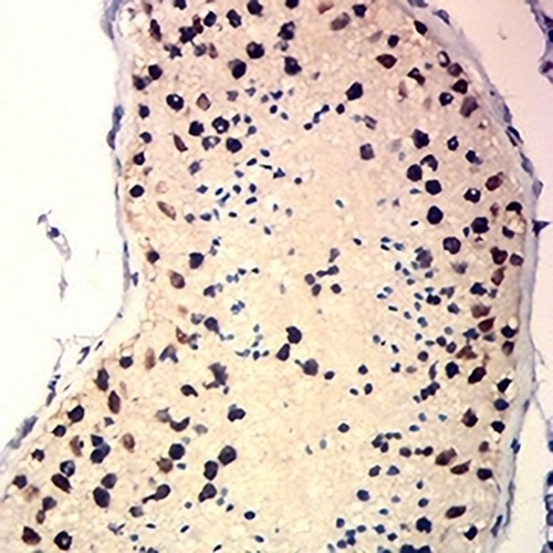
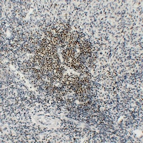
Immunohistochemical analysis of AKT (Phospho-Y315) staining in human brain formalin fixed paraffin embedded tissue section. The section was pre-treated using heat mediated antigen retrieval with sodium citrate buffer (pH 6.0). The section was then incubated with the antibody at room temperature and detected using an HRP conjugated compact polymer system. DAB was used as the chromogen. The section was then counterstained with haematoxylin and mounted with DPX.
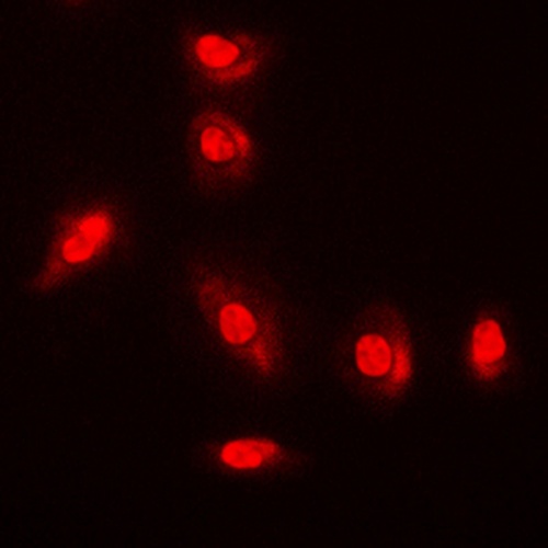
Immunofluorescent analysis of AKT (Phospho-Y315) staining in A549 cells. Formalin-fixed cells were permeabilized with 0.1% Triton X-100 in TBS for 5-10 minutes and blocked with 3% BSA-PBS for 30 minutes at room temperature. Cells were probed with the primary antibody in 3% BSA-PBS and incubated overnight at 4 °C in a humidified chamber. Cells were washed with PBST and incubated with a DyLight 594-conjugated secondary antibody (red) in PBS at room temperature in the dark.
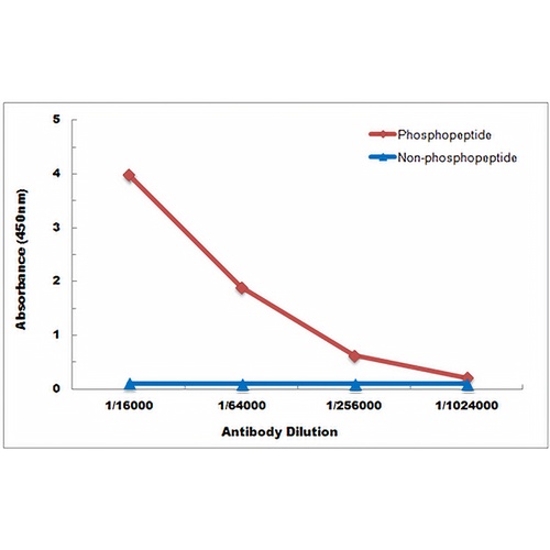
Direct ELISA antibody dose-response curve using Anti-AKT (Phospho-Y315) Antibody. Antigen (Phosphopeptide and non-phosphopeptide) concentration is 5 ug/ml. Goat Anti-Rabbit IgG (H&L) - HRP was used as the secondary antibody, and signal was developed by TMB substrate.
FAQs
 Neue Produkte