NCOA4 토끼 다클론항체
 데이터시트
데이터시트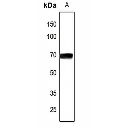
주요 기능 및 세부정보
- 대상:
- 소스/호스트:
- 반응성:
- 클론성:
- 신청:
- 활용:
- 저장:
-
브랜드:

 제품 세부정보
WB | 1:500 - 1:1000 |
IHC | 1:50 - 1:100 |
IF/ICC | 1:50 - 1:200 |
설명 | NCOA4에 대한 토끼 다클론 항체 |
특이성 | NCOA4 단백질의 내인성 수준을 인식합니다. |
항체 유형 | 1차 항체 |
면역원 | KLH-conjugated synthetic peptide encompassing a sequence within the center region of human NCOA4. The exact sequence is proprietary. |
정화 | 항체를 면역원 친화성 크로마토그래피로 정제하였다. |
분자량 | 예상: 32; 관찰됨: 70kD |
형태/버퍼 | Liquid in 0.42% Potassium phosphate, 0.87% Sodium chloride, pH 7.3, 30% glycerol, and 0.01% sodium azide. |
대체 이름 | ARA70; ELE1; RFG; Nuclear receptor coactivator 4; NCoA-4; Androgen receptor coactivator 70 kDa protein; 70 kDa AR-activator; 70 kDa androgen receptor coactivator; Androgen receptor-associated protein of 70 kDa; Ret-activating protein ELE1 |
유전자 기호 | NCOA4 |
엔트레즈 진 | 8031(인간) |
스위스프롯 | Q13772(인간) |
*Clone Number, Reactivity, Source/Host and Clonality can be found in the product name and Key Features section above.
Western blot analysis of NCOA4 expression in H1688 (A) whole cell lysates. (Predicted band size: 32; 69; 71; 73 kD; Observed band size: 70 kD)
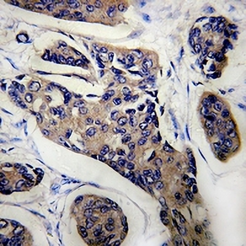
Immunohistochemical analysis of NCOA4 staining in human breast cancer formalin fixed paraffin embedded tissue section. The section was pre-treated using heat mediated antigen retrieval with sodium citrate buffer (pH 6.0). The section was then incubated with the antibody at room temperature and detected using an HRP conjugated compact polymer system. DAB was used as the chromogen. The section was then counterstained with haematoxylin and mounted with DPX.
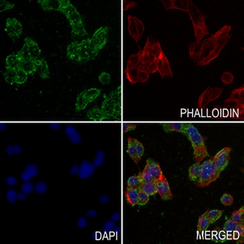
Immunofluorescent analysis of NCOA4 staining in MDAMB231 cells. Formalin-fixed cells were permeabilized with 0.1% Triton X-100 in TBS for 5-10 minutes and blocked with 3% BSA-PBS for 30 minutes at room temperature. Cells were probed with the primary antibody in 3% BSA-PBS and incubated overnight at 4 °C in a hidified chamber. Cells were washed with PBST and incubated with a AREX® Fluor 488 -conjugated secondary antibody (green) in PBS at room temperature in the dark. Phalloidin - AREX® Fluor 594 was used to stain Actin filaments (red). DAPI was used to stain the cell nuclei (blue).
자주 묻는 질문
 신제품
신제품