NMDAR2D Rabbit Polyclonal Antibody
 MSDS
MSDS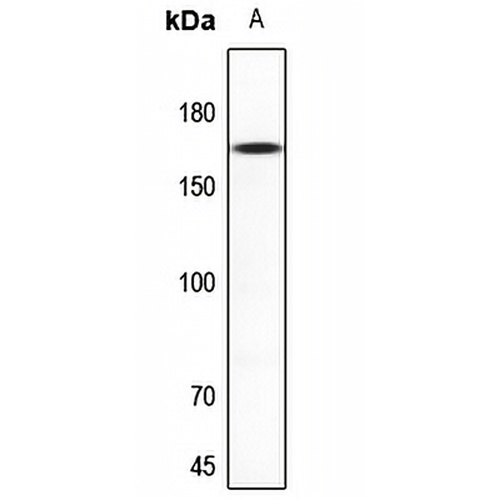
Key features and details
- Target:
- Source/Host:
- Reactivity:
- Clonality:
- Applications:
- Conjugation:
- Storage:
-
Brand:

 Product Details
WB | 1:500 - 1:1000 |
IF/ICC | 1:50 - 1:200 |
Description | Rabbit polyclonal antibody to NMDAR2D |
Specificity | Recognizes endogenous levels of NMDAR2D protein. |
Antibody Type | Primary antibody |
Imnunogen | KLH-conjugated synthetic peptide encompassing a sequence within the center region of human NMDAR2D. The exact sequence is proprietary. |
Purification | The antibody was purified by immunogen affinity chromatography. |
Molecular Weight | Predicted: 143 kD; Observed: 165 kD |
Form/Buffer | Liquid in 0.42% Potassium phosphate, 0.87% Sodium chloride, pH 7.3, 30% glycerol, and 0.01% sodium azide. |
Alternative Names | GluN2D; NMDAR2D; Glutamate receptor ionotropic, NMDA 2D; GluN2D; EB11; Glutamate [NMDA] receptor subunit epsilon-4; N-Methyl-D-aspartate receptor subtype 2D; NMDAR2D; NR2D |
Gene Symbol | GRIN2D |
Entrez Gene | 2906(Human); 14814(Mouse); 24412(Rat) |
SwissProt | O15399(Human); Q03391(Mouse); Q62645(Rat) |
*Clone Number, Reactivity, Source/Host and Clonality can be found in the product name and Key Features section above.
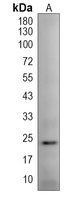
Western blot analysis of NMDAR2D expression in A549 (A) whole cell lysates. (Predicted band size: 143 kD; Observed band size: 165 kD)
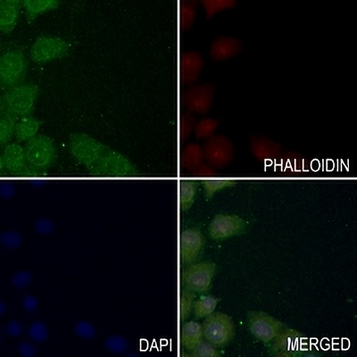
Immunofluorescent analysis of NMDAR2D staining in LO2 cells. Formalin-fixed cells were permeabilized with 0.1% Triton X-100 in TBS for 5-10 minutes and blocked with 3% BSA-PBS for 30 minutes at room temperature. Cells were probed with the primary antibody in 3% BSA-PBS and incubated overnight at 4 °C in a hidified chamber. Cells were washed with PBST and incubated with a AREX® Fluor 488 -conjugated secondary antibody (green) in PBS at room temperature in the dark. Phalloidin - AREX® Fluor 594 was used to stain Actin filaments (red). DAPI was used to stain the cell nuclei (blue).
FAQs
 New Products