Antibodi Poliklonal Kelinci Sitokrom P450 2B6
 MSDS
MSDS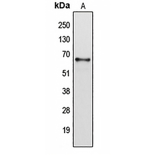
Fitur dan detail utama
- Sasaran:
- Sumber/Pembawa Acara:
- Reaktivitas:
- Klonalitas:
- Aplikasi:
- Konjugasi:
- Penyimpanan:
-
Merek:

 Detail Produk
WB | 1:500 - 1:1000 |
IHC | 1:50 - 1:200 |
JIKA/ICC | 1:50 - 1:200 |
Deskripsi | Antibodi poliklonal kelinci terhadap Sitokrom P450 2B6 |
Kekhususan | Mengenali kadar protein Sitokrom P450 2B6 endogen. |
Tipe Antibodi | Antibodi primer |
Imunogen | KLH-conjugated synthetic peptide encompassing a sequence within the center region of human Cytochrome P450 2B6. The exact sequence is proprietary. |
Pemurnian | Antibodi dimurnikan dengan kromatografi afinitas imunogen. |
Berat Molekul | Prediksi: 33; Teramati : 66 kD |
Formulir/Penyangga | Liquid in 0.42% Potassium phosphate, 0.87% Sodium chloride, pH 7.3, 30% glycerol, and 0.01% sodium azide. |
Nama Alternatif | Cytochrome P450 2B6; 1,4-cineole 2-exo-monooxygenase; CYPIIB6; Cytochrome P450 IIB1 |
Simbol Gen | CYP2B6 |
Entrez Gene | 1555 (Manusia) |
SwissProt | P20813(Manusia) |
*Clone Number, Reactivity, Source/Host and Clonality can be found in the product name and Key Features section above.
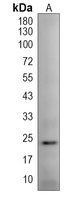
Western blot analysis of Cytochrome P450 2B6 expression in HepG2 (A) whole cell lysates. (Predicted band size: 33; 56 kD; Observed band size: 66 kD)
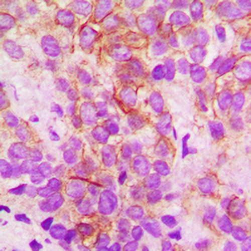
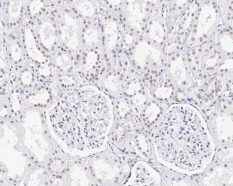
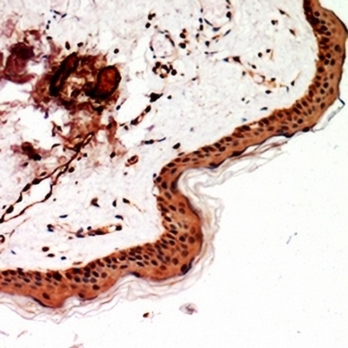
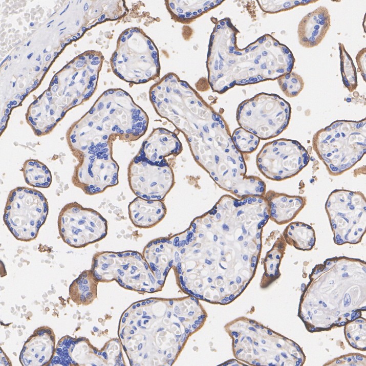
Immunohistochemical analysis of Cytochrome P450 2B6 staining in human breast cancer formalin fixed paraffin embedded tissue section. The section was pre-treated using heat mediated antigen retrieval with sodium citrate buffer (pH 6.0). The section was then incubated with the antibody at room temperature and detected using an HRP conjugated compact polymer system. DAB was used as the chromogen. The section was then counterstained with haematoxylin and mounted with DPX.
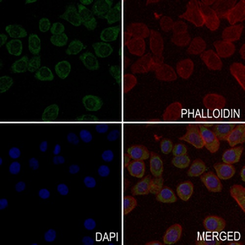
Immunofluorescent analysis of Cytochrome P450 2B6 staining in PCCL cells. Formalin-fixed cells were permeabilized with 0.1% Triton X-100 in TBS for 5-10 minutes and blocked with 3% BSA-PBS for 30 minutes at room temperature. Cells were probed with the primary antibody in 3% BSA-PBS and incubated overnight at 4 °C in a hidified chamber. Cells were washed with PBST and incubated with a AREX® Fluor 488 -conjugated secondary antibody (green) in PBS at room temperature in the dark. Phalloidin - AREX® Fluor 594 was used to stain Actin filaments (red). DAPI was used to stain the cell nuclei (blue).
FAQ
 Produk Baru