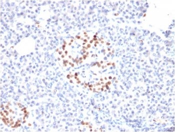
Antibodi Monoklonal Tikus AKT (C2501)
 MSDS
MSDS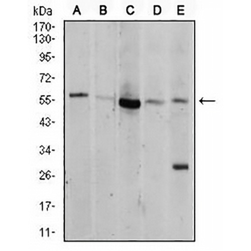
Fitur dan detail utama
- Sasaran:
- Sumber/Pembawa Acara:
- Reaktivitas:
- Klonalitas:
- Aplikasi:
- Konjugasi:
- Penyimpanan:
-
Merek:

 Detail Produk
WB | 1:500 - 1:1000 |
JIKA/ICC | 1:100 - 1:500 |
FC | 1:100 - 1:200 |
Deskripsi | Mouse monoklonal ke AKT |
Kekhususan | Mengenali kadar protein AKT endogen |
Tipe Antibodi | Antibodi primer |
Imunogen | Recombinant fusion protein of human AKT expressed in E. Coli |
Pemurnian | This antibody is purified through a protein G column. |
Berat Molekul | Prediksi: 56 kD; Teramati : 55 kD kD |
Formulir/Penyangga | Mouse IgG1. Liquid in PBS, pH 7.3, 30% glycerol, and 0.01% sodium azide. |
Nama Alternatif | PKB; RAC; RAC-alpha serine/threonine-protein kinase; Protein kinase B; PKB; Protein kinase B alpha; PKB alpha; Proto-oncogene c-Akt; RAC-PK-alpha |
Simbol Gen | AKT1 |
Entrez Gene | 207(Manusia); 11651(Tikus); 24185 (Tikus) |
SwissProt | P31749(Manusia); P31750(Tikus); P47196(Tikus) |
*Clone Number, Reactivity, Source/Host and Clonality can be found in the product name and Key Features section above.
Western blot analysis of AKT expression in MCF7 (A), NIH/3T3 (B), Hela (C), COS7 (D), C6 (E) whole cell lysates. (Predicted band size: 56 kD; Observed band size: 55 kD kD)
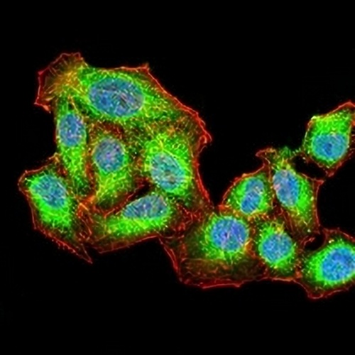
Immunofluorescent analysis of AKT staining in HepG2 cells. Formalin-fixed cells were permeabilized with 0.1% Triton X-100 in TBS for 5-10 minutes and blocked with 3% BSA-PBS for 30 minutes at room temperature. Cells were probed with the primary antibody in 3% BSA-PBS and incubated overnight at 4 °C in a hidified chamber. Cells were washed with PBST and incubated with an AREX® Fluor 488 -conjugated secondary antibody (green) in PBS at room temperature in the dark. Phalloidin - AREX® Fluor 594 was used to stain Actin filaments (red). DAPI was used to stain the cell nuclei (blue).
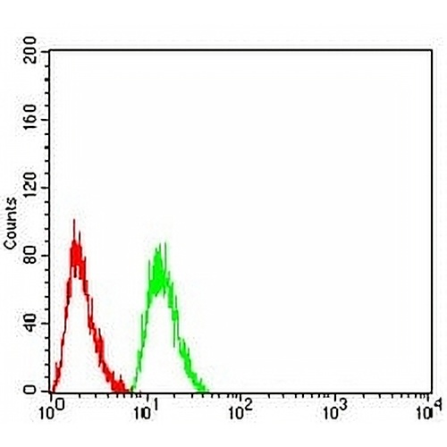
Flow cytometric analysis of HeLa cells using Anti-AKT Antibody (green) and negative control (red).
FAQ
 Produk Baru