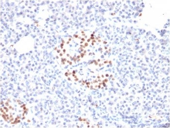
MAP2 Mouse Monoclonal Antibody(C3661)
 Sicherheitsdatenblatt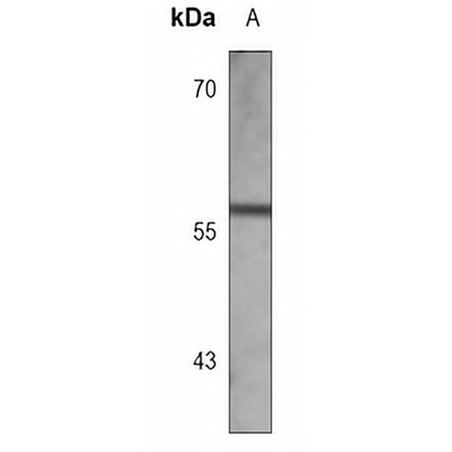
Hauptmerkmale und Details
- Ziel:
- Quelle/Host:
- Reaktivität:
- Klonalität:
- Anwendungen:
- Konjugation:
- Lagerung:
-
Marke:

 Produktdetails
WB | 1:500 - 1:1000 |
IHC | 1:50 - 1:200 |
IF/ICC | 1:10 - 1:50 |
Beschreibung | Mouse monoclonal antibody to MAP2 |
Spezifität | Recognizes endogenous levels of MAP2 protein. |
Antikörpertyp | Primärer Antikörper |
Immunogen | Recombinant fusion protein of human MAP2. The exact sequence is proprietary. |
Reinigung | |
Molekulargewicht | Predicted: 199 kD; Observed: 56 kD |
Form/Puffer | Mouse IgG1 kappa. Supplied in crude ascites with 0.01% sodium azide. |
Alternative Namen | Microtubule-associated protein 2; MAP-2 |
Gensymbol | KARTE2 |
Entrez Gene | 4133 (Mensch) |
SwissProt | P11137 (Mensch) |
*Clone Number, Reactivity, Source/Host and Clonality can be found in the product name and Key Features section above.
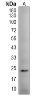
Western blot analysis of MAP2 expression in MCF7 (A) whole cell lysates. (Predicted band size: 199 kD; Observed band size: 56 kD)
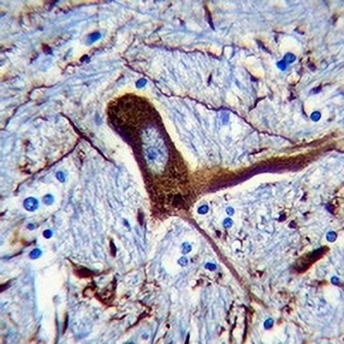
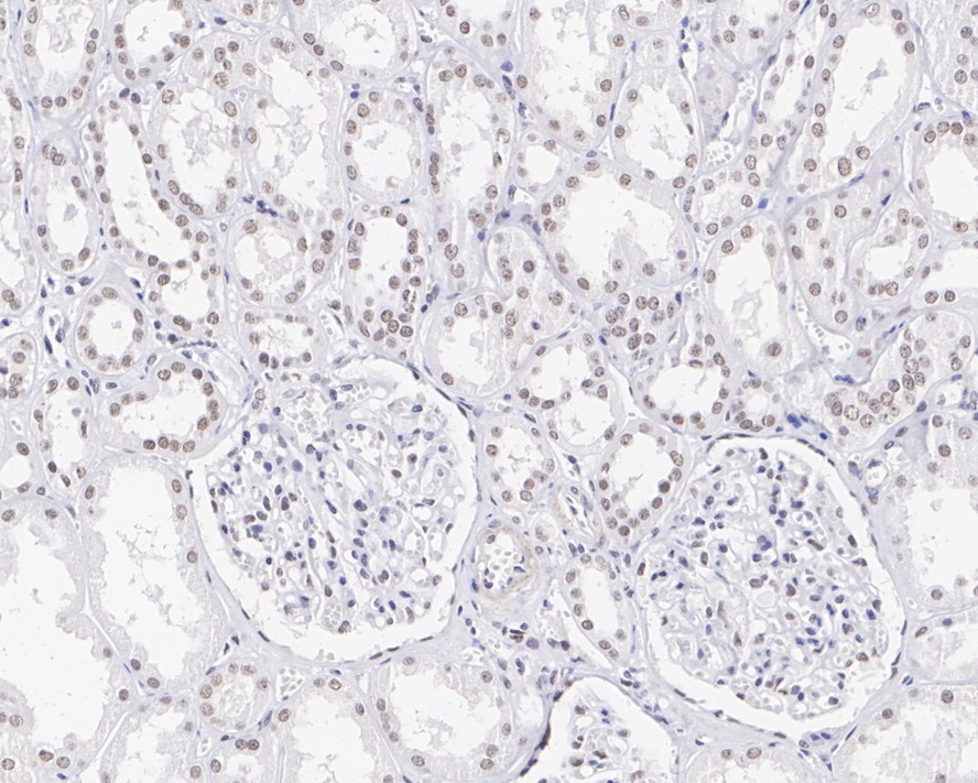
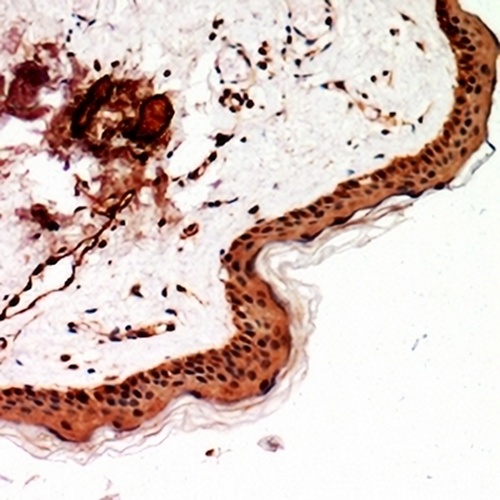
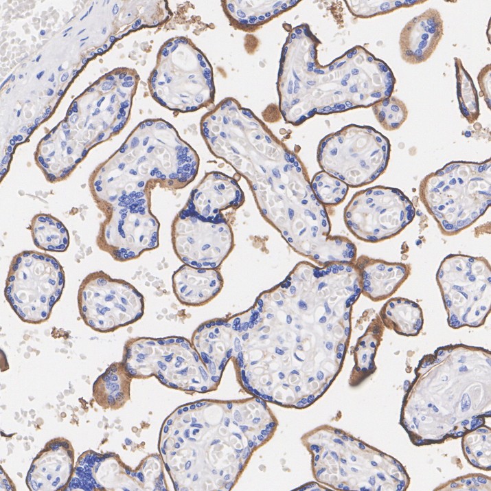
Immunohistochemical analysis of MAP2 staining in human brain formalin fixed paraffin embedded tissue section. The section was pre-treated using heat mediated antigen retrieval with sodium citrate buffer (pH 6.0). The section was then incubated with the antibody at room temperature and detected using an HRP conjugated compact polymer system. DAB was used as the chromogen. The section was then counterstained with haematoxylin and mounted with DPX.
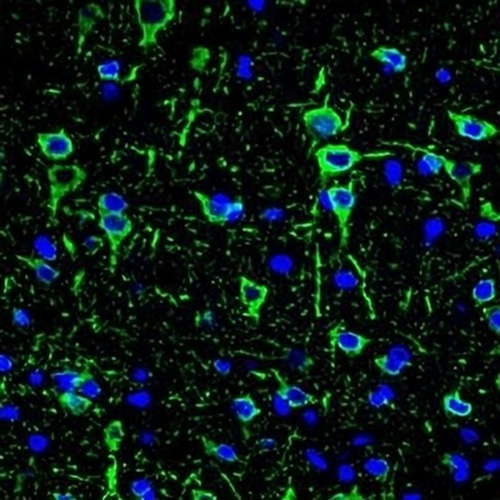
Immunofluorescent analysis of MAP2 staining in brain tissue cells. Formalin-fixed cells were permeabilized with 0.1% Triton X-100 in TBS for 5-10 minutes and blocked with 3% BSA-PBS for 30 minutes at room temperature. Cells were probed with the primary antibody in 3% BSA-PBS and incubated overnight at 4 °C in a humidified chamber. Cells were washed with PBST and incubated with a AREX® Fluor 488 -conjugated secondary antibody (green) in PBS at room temperature in the dark. DAPI was used to stain the cell nuclei (blue).
FAQs
 Neue Produkte